Endo180 Polyclonal Antibody
- Catalog No.:YT1556
- Applications:WB;IHC;IF;ELISA
- Reactivity:Human;Mouse;Rat
- Target:
- Endo180
- Fields:
- >>Phagosome;>>Tuberculosis
- Gene Name:
- MRC2
- Protein Name:
- C-type mannose receptor 2
- Human Gene Id:
- 9902
- Human Swiss Prot No:
- Q9UBG0
- Mouse Gene Id:
- 17534
- Mouse Swiss Prot No:
- Q64449
- Rat Gene Id:
- 498011
- Rat Swiss Prot No:
- Q4TU93
- Immunogen:
- The antiserum was produced against synthesized peptide derived from human MRC2. AA range:121-170
- Specificity:
- Endo180 Polyclonal Antibody detects endogenous levels of Endo180 protein.
- Formulation:
- Liquid in PBS containing 50% glycerol, 0.5% BSA and 0.02% sodium azide.
- Source:
- Polyclonal, Rabbit,IgG
- Dilution:
- WB 1:500 - 1:2000. IHC 1:100 - 1:300. IF 1:200 - 1:1000. ELISA: 1:20000. Not yet tested in other applications.
- Purification:
- The antibody was affinity-purified from rabbit antiserum by affinity-chromatography using epitope-specific immunogen.
- Concentration:
- 1 mg/ml
- Storage Stability:
- -15°C to -25°C/1 year(Do not lower than -25°C)
- Other Name:
- MRC2;CLEC13E;ENDO180;KIAA0709;UPARAP;C-type mannose receptor 2;C-type lectin domain family 13 member E;Endocytic receptor 180;Macrophage mannose receptor 2;Urokinase-type plasminogen activator receptor-associated protein;UPAR-asso
- Observed Band(KD):
- 167kD
- Background:
- mannose receptor C type 2(MRC2) Homo sapiens This gene encodes a member of the mannose receptor family of proteins that contain a fibronectin type II domain and multiple C-type lectin-like domains. The encoded protein plays a role in extracellular matrix remodeling by mediating the internalization and lysosomal degradation of collagen ligands. Expression of this gene may play a role in the tumorigenesis and metastasis of several malignancies including breast cancer, gliomas and metastatic bone disease. [provided by RefSeq, Feb 2012],
- Function:
- domain:C-type lectin domains 3 to 8 are not required for calcium-dependent binding of mannose, fucose and N-acetylglucosamine. C-type lectin domain 2 is responsible for sugar-binding in a calcium-dependent manner.,domain:Fibronectin type-II domain mediates collagen-binding.,domain:Ricin B-type lectin domain contacts with the second C-type lectin domain.,function:May play a role as endocytotic lectin receptor displaying calcium-dependent lectin activity. Internalizes glycosylated ligands from the extracellular space for release in an endosomal compartment via clathrin-mediated endocytosis. May be involved in plasminogen activation system controlling the extracellular level of PLAUR/PLAU, and thus may regulate protease activity at the cell surface. May contribute to cellular uptake, remodeling and degradation of extracellular collagen matrices. May play a role during cancer progression as
- Subcellular Location:
- Membrane; Single-pass type I membrane protein.
- Expression:
- Ubiquitous with low expression in brain, placenta, lung, kidney, pancreas, spleen, thymus and colon. Expressed in endothelial cells, fibroblasts and macrophages. Highly expressed in fetal lung and kidney.
- June 19-2018
- WESTERN IMMUNOBLOTTING PROTOCOL
- June 19-2018
- IMMUNOHISTOCHEMISTRY-PARAFFIN PROTOCOL
- June 19-2018
- IMMUNOFLUORESCENCE PROTOCOL
- September 08-2020
- FLOW-CYTOMEYRT-PROTOCOL
- May 20-2022
- Cell-Based ELISA│解您多样本WB检测之困扰
- July 13-2018
- CELL-BASED-ELISA-PROTOCOL-FOR-ACETYL-PROTEIN
- July 13-2018
- CELL-BASED-ELISA-PROTOCOL-FOR-PHOSPHO-PROTEIN
- July 13-2018
- Antibody-FAQs
- Products Images

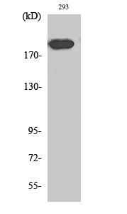
- Western Blot analysis of various cells using Endo180 Polyclonal Antibody diluted at 1:1000

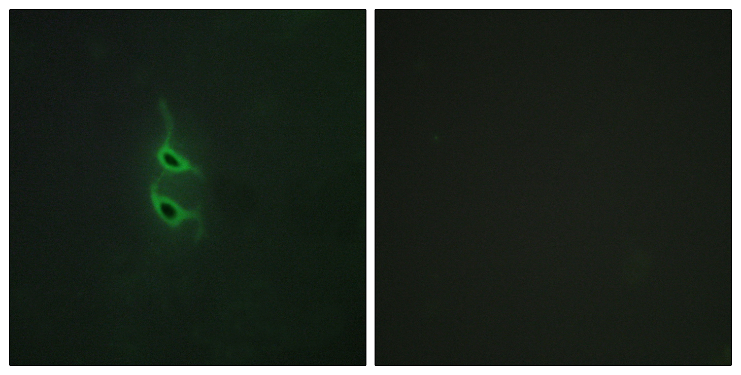
- Immunofluorescence analysis of HepG2 cells, using MRC2 Antibody. The picture on the right is blocked with the synthesized peptide.

- Immunohistochemistry analysis of paraffin-embedded human brain tissue, using MRC2 Antibody. The picture on the right is blocked with the synthesized peptide.

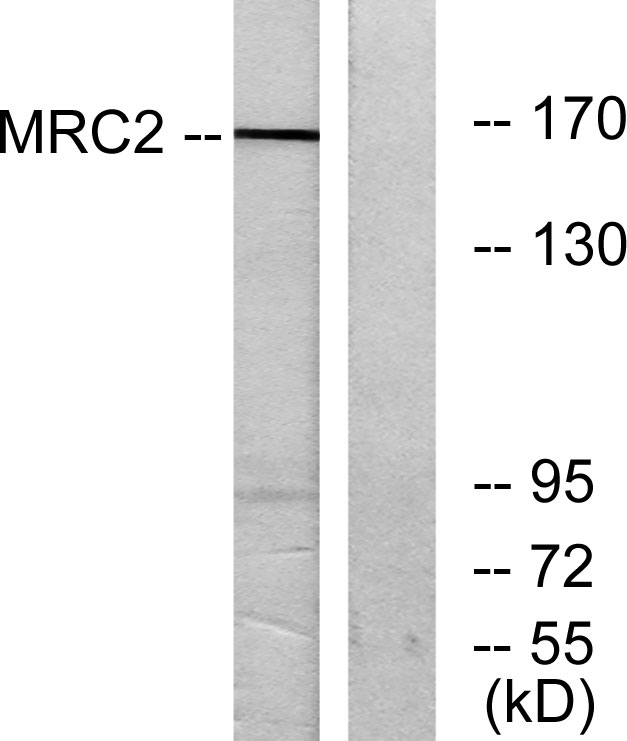
- Western blot analysis of lysates from 293 cells, using MRC2 Antibody. The lane on the right is blocked with the synthesized peptide.

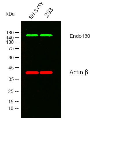
- Western blot analysis of lysates from SH-SY5Y,293 cells, (Green) primary antibody was diluted at 1:1000, 4°over night, secondary antibody was diluted at 1:10000, 37° 1hour. (Red) loading contrl antibody was diluted at 1:5000 as loading control, 4° over night,secondary antibody was diluted at 1:10000, 37° 1hour.


