FAS Polyclonal Antibody
- Catalog No.:YT1676
- Applications:WB;IHC;IF;ELISA
- Reactivity:Human;Rat;Mouse;Canine
- Target:
- FAS
- Fields:
- >>Platinum drug resistance;>>MAPK signaling pathway;>>Cytokine-cytokine receptor interaction;>>p53 signaling pathway;>>Apoptosis;>>Necroptosis;>>Natural killer cell mediated cytotoxicity;>>TNF signaling pathway;>>Non-alcoholic fatty liver disease;>>Alcoholic liver disease;>>Type I diabetes mellitus;>>Alzheimer disease;>>Pathways of neurodegeneration - multiple diseases;>>Pathogenic Escherichia coli infection;>>Chagas disease;>>African trypanosomiasis;>>Hepatitis C;>>Hepatitis B;>>Measles;>>Human cytomegalovirus infection;>>Influenza A;>>Human papillomavirus infection;>>Kaposi sarcoma-associated herpesvirus infection;>>Herpes simplex virus 1 infection;>>Epstein-Barr virus infection;>>Human immunodeficiency virus 1 infection;>>Pathways in cancer;>>Proteoglycans in cancer;>>Autoimmune thyroid disease;>>Allograft rejection;>>Graft-versus-host disease;>>Lipid and atherosclerosis
- Gene Name:
- FAS
- Protein Name:
- Tumor necrosis factor receptor superfamily member 6
- Human Gene Id:
- 355
- Human Swiss Prot No:
- P25445
- Mouse Swiss Prot No:
- P25446
- Immunogen:
- The antiserum was produced against synthesized peptide derived from human FAS. AA range:257-306
- Specificity:
- FAS Polyclonal Antibody detects endogenous levels of FAS protein.
- Formulation:
- Liquid in PBS containing 50% glycerol, 0.5% BSA and 0.02% sodium azide.
- Source:
- Polyclonal, Rabbit,IgG
- Dilution:
- WB 1:500-2000, IF 1:50-300, IHC 1:50-300
- Purification:
- The antibody was affinity-purified from rabbit antiserum by affinity-chromatography using epitope-specific immunogen.
- Concentration:
- 1 mg/ml
- Storage Stability:
- -15°C to -25°C/1 year(Do not lower than -25°C)
- Other Name:
- FAS;APT1;FAS1;TNFRSF6;Tumor necrosis factor receptor superfamily member 6;Apo-1 antigen;Apoptosis-mediating surface antigen FAS;FASLG receptor;CD antigen CD95
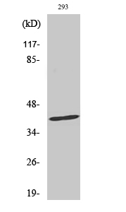
- Observed Band(KD):
- 42kD
- Background:
- The protein encoded by this gene is a member of the TNF-receptor superfamily. This receptor contains a death domain. It has been shown to play a central role in the physiological regulation of programmed cell death, and has been implicated in the pathogenesis of various malignancies and diseases of the immune system. The interaction of this receptor with its ligand allows the formation of a death-inducing signaling complex that includes Fas-associated death domain protein (FADD), caspase 8, and caspase 10. The autoproteolytic processing of the caspases in the complex triggers a downstream caspase cascade, and leads to apoptosis. This receptor has been also shown to activate NF-kappaB, MAPK3/ERK1, and MAPK8/JNK, and is found to be involved in transducing the proliferating signals in normal diploid fibroblast and T cells. Several alternatively spliced transcript variants have been described, s
- Function:
- disease:Defects in FAS are the cause of autoimmune lymphoproliferative syndrome type 1A (ALPS1A) [MIM:601859]; also known as Canale-Smith syndrome (CSS). ALPS is a childhood syndrome involving hemolytic anemia and thrombocytopenia with massive lymphadenopathy and splenomegaly.,domain:Contains a death domain involved in the binding of FADD, and maybe to other cytosolic adapter proteins.,function:Receptor for TNFSF6/FASLG. The adapter molecule FADD recruits caspase-8 to the activated receptor. The resulting death-inducing signaling complex (DISC) performs caspase-8 proteolytic activation which initiates the subsequent cascade of caspases (aspartate-specific cysteine proteases) mediating apoptosis. FAS-mediated apoptosis may have a role in the induction of peripheral tolerance, in the antigen-stimulated suicide of mature T-cells, or both. The secreted isoforms 2 to 6 block apoptosis (in vit
- Subcellular Location:
- [Isoform 1]: Cell membrane ; Single-pass type I membrane protein . Membrane raft .; [Isoform 2]: Secreted.; [Isoform 3]: Secreted.; [Isoform 4]: Secreted.; [Isoform 5]: Secreted.; [Isoform 6]: Secreted.
- Expression:
- Isoform 1 and isoform 6 are expressed at equal levels in resting peripheral blood mononuclear cells. After activation there is an increase in isoform 1 and decrease in the levels of isoform 6.
Anti-apoptotic and apoptotic pathway analysis of arsenic trioxide‑induced apoptosis in human gastric cancer SGC-7901 cells. ONCOLOGY REPORTS 2014 Jun 20 WB Human 1:800 SGC-7901 cell
Protective Effect of Adipose-Derived Mesenchymal Stem Cell Secretome against Hepatocyte Apoptosis Induced by Liver Ischemia-Reperfusion with Partial Hepatectomy Injury. Stem Cells International Stem Cells Int. 2021;2021:9969372 IHC Pig Liver
The preparation of a cold-water soluble polysaccharide from Grifola frondosa and its inhibitory effects on MKN-45 cells. GLYCOCONJUGATE JOURNAL 2020 Jun 15 WB Human MKN-45 cell
Fucoidan inhibits proliferation of the SKM-1 acute myeloid leukaemia cell line via the activation of apoptotic pathways and production of reactive oxygen species. Molecular Medicine Reports Mol Med Rep. 2015 Nov;12(5):6649-6655 WB Human SKM-1 cell
- June 19-2018
- WESTERN IMMUNOBLOTTING PROTOCOL
- June 19-2018
- IMMUNOHISTOCHEMISTRY-PARAFFIN PROTOCOL
- June 19-2018
- IMMUNOFLUORESCENCE PROTOCOL
- September 08-2020
- FLOW-CYTOMEYRT-PROTOCOL
- May 20-2022
- Cell-Based ELISA│解您多样本WB检测之困扰
- July 13-2018
- CELL-BASED-ELISA-PROTOCOL-FOR-ACETYL-PROTEIN
- July 13-2018
- CELL-BASED-ELISA-PROTOCOL-FOR-PHOSPHO-PROTEIN
- July 13-2018
- Antibody-FAQs
- Products Images
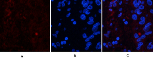
- Immunofluorescence analysis of human-liver-cancer tissue. 1,FAS Polyclonal Antibody(red) was diluted at 1:200(4°C,overnight). 2, Cy3 labled Secondary antibody was diluted at 1:300(room temperature, 50min).3, Picture B: DAPI(blue) 10min. Picture A:Target. Picture B: DAPI. Picture C: merge of A+B
- Immunofluorescence analysis of human-liver-cancer tissue. 1,FAS Polyclonal Antibody(red) was diluted at 1:200(4°C,overnight). 2, Cy3 labled Secondary antibody was diluted at 1:300(room temperature, 50min).3, Picture B: DAPI(blue) 10min. Picture A:Target. Picture B: DAPI. Picture C: merge of A+B
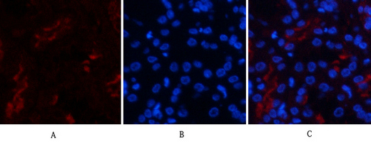
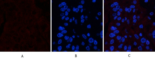
- Immunofluorescence analysis of human-kidney tissue. 1,FAS Polyclonal Antibody(red) was diluted at 1:200(4°C,overnight). 2, Cy3 labled Secondary antibody was diluted at 1:300(room temperature, 50min).3, Picture B: DAPI(blue) 10min. Picture A:Target. Picture B: DAPI. Picture C: merge of A+B

- Immunofluorescence analysis of human-kidney tissue. 1,FAS Polyclonal Antibody(red) was diluted at 1:200(4°C,overnight). 2, Cy3 labled Secondary antibody was diluted at 1:300(room temperature, 50min).3, Picture B: DAPI(blue) 10min. Picture A:Target. Picture B: DAPI. Picture C: merge of A+B
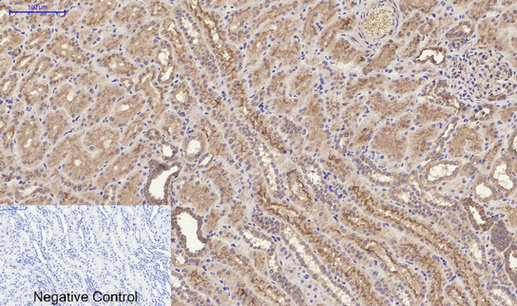
- Immunohistochemical analysis of paraffin-embedded Human-kidney tissue. 1,FAS Polyclonal Antibody was diluted at 1:200(4°C,overnight). 2, Sodium citrate pH 6.0 was used for antibody retrieval(>98°C,20min). 3,Secondary antibody was diluted at 1:200(room tempeRature, 30min). Negative control was used by secondary antibody only.
- Western Blot analysis of various cells using FAS Polyclonal Antibody

- Immunohistochemical analysis of paraffin-embedded Human testis. 1, Antibody was diluted at 1:100(4° overnight). 2, High-pressure and temperature EDTA, pH8.0 was used for antigen retrieval. 3,Secondary antibody was diluted at 1:200(room temperature, 30min).
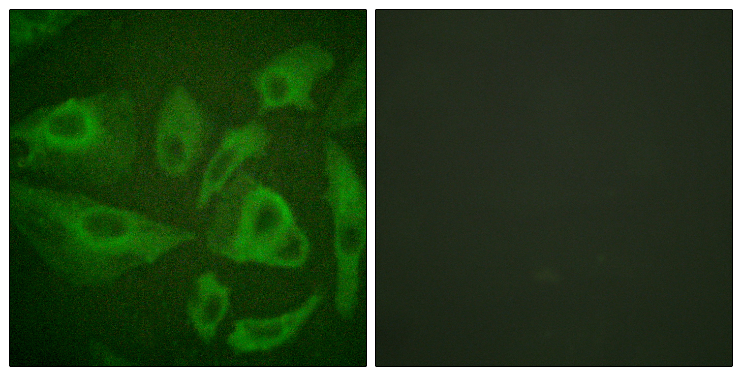
- Immunofluorescence analysis of HeLa cells, using FAS Antibody. The picture on the right is blocked with the synthesized peptide.
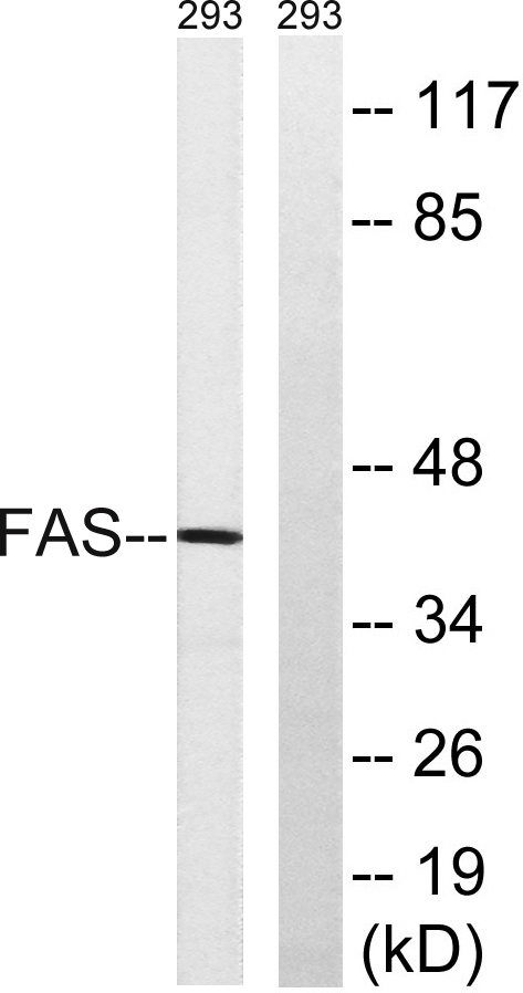
- Western blot analysis of lysates from 293 cells, using FAS Antibody. The lane on the right is blocked with the synthesized peptide.


