FGF Receptor 1 Polyclonal Antibody
- Catalog No.:YT1716
- Applications:WB;IF;ELISA
- Reactivity:Human;Mouse;Rat
- Target:
- FGF Receptor 1
- Fields:
- >>MAPK signaling pathway;>>Ras signaling pathway;>>Rap1 signaling pathway;>>Calcium signaling pathway;>>PI3K-Akt signaling pathway;>>Adherens junction;>>Signaling pathways regulating pluripotency of stem cells;>>Thermogenesis;>>Regulation of actin cytoskeleton;>>Parathyroid hormone synthesis, secretion and action;>>Pathways in cancer;>>Proteoglycans in cancer;>>Prostate cancer;>>Melanoma;>>Breast cancer;>>Central carbon metabolism in cancer
- Gene Name:
- FGFR1 BFGFR CEK FGFBR FLG FLT2 HBGFR
- Protein Name:
- Fibroblast growth factor receptor 1
- Human Gene Id:
- 2260
- Human Swiss Prot No:
- P11362
- Mouse Gene Id:
- 14182
- Mouse Swiss Prot No:
- P16092
- Rat Gene Id:
- 79114
- Rat Swiss Prot No:
- Q04589
- Immunogen:
- The antiserum was produced against synthesized peptide derived from human FGFR1. AA range:626-675
- Specificity:
- Flg Polyclonal Antibody detects endogenous levels of Flg protein.
- Formulation:
- Liquid in PBS containing 50% glycerol, 0.5% BSA and 0.02% sodium azide.
- Source:
- Polyclonal, Rabbit,IgG
- Dilution:
- WB 1:500 - 1:2000. IF 1:200 - 1:1000. ELISA: 1:5000. Not yet tested in other applications.
- Purification:
- The antibody was affinity-purified from rabbit antiserum by affinity-chromatography using epitope-specific immunogen.
- Concentration:
- 1 mg/ml
- Storage Stability:
- -15°C to -25°C/1 year(Do not lower than -25°C)
- Other Name:
- FGFR1;BFGFR;CEK;FGFBR;FLG;FLT2;HBGFR;Fibroblast growth factor receptor 1;FGFR-1;Basic fibroblast growth factor receptor 1;BFGFR;bFGF-R-1;Fms-like tyrosine kinase 2;FLT-2;N-sam;Proto-oncogene c-Fgr;CD antigen CD331
- Molecular Weight(Da):
- 120kD
- Observed Band(KD):
- full length 120-140kD,FOP-FGFR1 90kD
- Background:
- The protein encoded by this gene is a member of the fibroblast growth factor receptor (FGFR) family, where amino acid sequence is highly conserved between members and throughout evolution. FGFR family members differ from one another in their ligand affinities and tissue distribution. A full-length representative protein consists of an extracellular region, composed of three immunoglobulin-like domains, a single hydrophobic membrane-spanning segment and a cytoplasmic tyrosine kinase domain. The extracellular portion of the protein interacts with fibroblast growth factors, setting in motion a cascade of downstream signals, ultimately influencing mitogenesis and differentiation. This particular family member binds both acidic and basic fibroblast growth factors and is involved in limb induction. Mutations in this gene have been associated with Pfeiffer syndrome, Jackson-Weiss syndrome,
- Function:
- catalytic activity:ATP + a [protein]-L-tyrosine = ADP + a [protein]-L-tyrosine phosphate.,disease:A chromosomal aberration involving FGFR1 may be a cause of stem cell leukemia lymphoma syndrome (SCLL). Translocation t(8;13)(p11;q12) with ZMYM2. SCLL usually presents as lymphoblastic lymphoma in association with a myeloproliferative disorder, often accompanied by pronounced peripheral eosinophilia and/or prominent eosinophilic infiltrates in the affected bone marrow.,disease:A chromosomal aberration involving FGFR1 may be a cause of stem cell myeloproliferative disorder (MPD). Translocation t(6;8)(q27;p11) with FGFR1OP. Insertion ins(12;8)(p11;p11p22) with FGFR1OP2. MPD is characterized by myeloid hyperplasia, eosinophilia and T-cell or B-cell lymphoblastic lymphoma. In general it progresses to acute myeloid leukemia. The fusion proteins FGFR1OP2-FGFR1, FGFR1OP-FGFR1 or FGFR1-FGFR1OP may
- Subcellular Location:
- Cell membrane; Single-pass type I membrane protein. Nucleus. Cytoplasm, cytosol. Cytoplasmic vesicle. After ligand binding, both receptor and ligand are rapidly internalized. Can translocate to the nucleus after internalization, or by translocation from the endoplasmic reticulum or Golgi apparatus to the cytosol, and from there to the nucleus.
- Expression:
- Detected in astrocytoma, neuroblastoma and adrenal cortex cell lines. Some isoforms are detected in foreskin fibroblast cell lines, however isoform 17, isoform 18 and isoform 19 are not detected in these cells.
- June 19-2018
- WESTERN IMMUNOBLOTTING PROTOCOL
- June 19-2018
- IMMUNOHISTOCHEMISTRY-PARAFFIN PROTOCOL
- June 19-2018
- IMMUNOFLUORESCENCE PROTOCOL
- September 08-2020
- FLOW-CYTOMEYRT-PROTOCOL
- May 20-2022
- Cell-Based ELISA│解您多样本WB检测之困扰
- July 13-2018
- CELL-BASED-ELISA-PROTOCOL-FOR-ACETYL-PROTEIN
- July 13-2018
- CELL-BASED-ELISA-PROTOCOL-FOR-PHOSPHO-PROTEIN
- July 13-2018
- Antibody-FAQs
- Products Images

- Western Blot analysis of various cells using Flg Polyclonal Antibody diluted at 1:1000

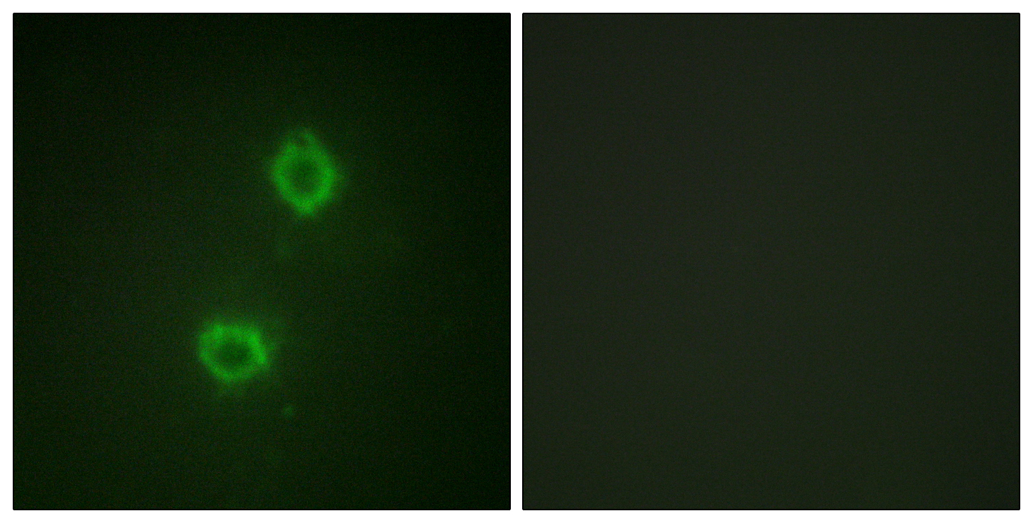
- Immunofluorescence analysis of HUVEC cells, using FGFR1 Antibody. The picture on the right is blocked with the synthesized peptide.

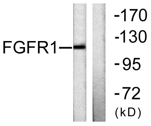
- Western blot analysis of lysates from 293 cells, using FGFR1 Antibody. The lane on the right is blocked with the synthesized peptide.

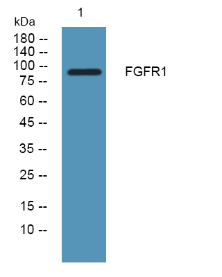
- Western blot analysis of lysates from SH-SY5Y cells, primary antibody was diluted at 1:1000, 4°over night


