Gα t2 Polyclonal Antibody
- Catalog No.:YT2095
- Applications:WB;IHC;IF;ELISA
- Reactivity:Human;Mouse
- Target:
- Gα t2
- Fields:
- >>Phototransduction
- Gene Name:
- GNAT2
- Protein Name:
- Guanine nucleotide-binding protein G(t) subunit alpha-2
- Human Gene Id:
- 2780
- Human Swiss Prot No:
- P19087
- Mouse Gene Id:
- 14686
- Mouse Swiss Prot No:
- P50149
- Immunogen:
- The antiserum was produced against synthesized peptide derived from human GNAT2. AA range:1-50
- Specificity:
- Gα t2 Polyclonal Antibody detects endogenous levels of Gα t2 protein.
- Formulation:
- Liquid in PBS containing 50% glycerol, 0.5% BSA and 0.02% sodium azide.
- Source:
- Polyclonal, Rabbit,IgG
- Dilution:
- WB 1:500 - 1:2000. IHC 1:100 - 1:300. ELISA: 1:10000.. IF 1:50-200
- Purification:
- The antibody was affinity-purified from rabbit antiserum by affinity-chromatography using epitope-specific immunogen.
- Concentration:
- 1 mg/ml
- Storage Stability:
- -15°C to -25°C/1 year(Do not lower than -25°C)
- Other Name:
- GNAT2;GNATC;Guanine nucleotide-binding protein G(t) subunit alpha-2;Transducin alpha-2 chain
- Observed Band(KD):
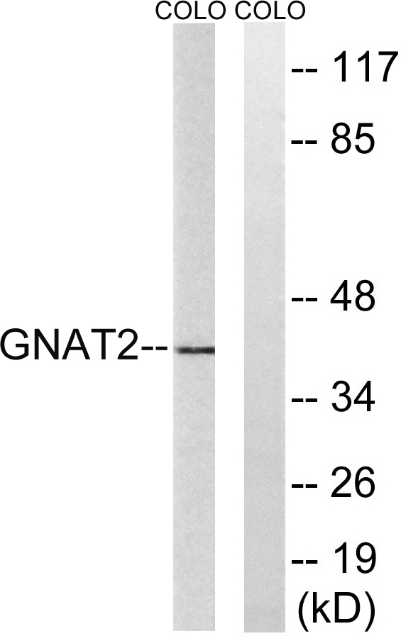
- 40kD
- Background:
- Transducin is a 3-subunit guanine nucleotide-binding protein (G protein) which stimulates the coupling of rhodopsin and cGMP-phoshodiesterase during visual impulses. The transducin alpha subunits in rods and cones are encoded by separate genes. This gene encodes the alpha subunit in cones. [provided by RefSeq, Jul 2008],
- Function:
- disease:Defects in GNAT2 are the cause of achromatopsia type 4 (ACHM4) [MIM:139340]. Achromatopsia is an autosomal recessively inherited visual disorder that is present from birth and that features the absence of color discrimination.,function:Guanine nucleotide-binding proteins (G proteins) are involved as modulators or transducers in various transmembrane signaling systems. Transducin is an amplifier and one of the transducers of a visual impulse that performs the coupling between rhodopsin and cGMP-phosphodiesterase.,similarity:Belongs to the G-alpha family. G(i/o/t/z) subfamily.,subunit:G proteins are composed of 3 units; alpha, beta and gamma. The alpha chain contains the guanine nucleotide binding site.,tissue specificity:Retinal rod outer segment.,
- Subcellular Location:
- Cell projection, cilium, photoreceptor outer segment . Photoreceptor inner segment . Localizes mainly in the outer segment in the dark-adapted state, whereas is translocated to the inner part of the photoreceptors in the light-adapted state. During dark-adapted conditions, in the presence of UNC119 mislocalizes from the outer segment to the inner part of rod photoreceptors which leads to decreased photoreceptor damage caused by light. .
- Expression:
- Retinal rod outer segment.
- June 19-2018
- WESTERN IMMUNOBLOTTING PROTOCOL
- June 19-2018
- IMMUNOHISTOCHEMISTRY-PARAFFIN PROTOCOL
- June 19-2018
- IMMUNOFLUORESCENCE PROTOCOL
- September 08-2020
- FLOW-CYTOMEYRT-PROTOCOL
- May 20-2022
- Cell-Based ELISA│解您多样本WB检测之困扰
- July 13-2018
- CELL-BASED-ELISA-PROTOCOL-FOR-ACETYL-PROTEIN
- July 13-2018
- CELL-BASED-ELISA-PROTOCOL-FOR-PHOSPHO-PROTEIN
- July 13-2018
- Antibody-FAQs
- Products Images

- Western blot analysis of lysates from COLO cells, using GNAT2 Antibody. The lane on the right is blocked with the synthesized peptide.

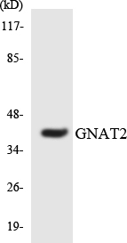
- Western blot analysis of the lysates from Jurkat cells using GNAT2 antibody.

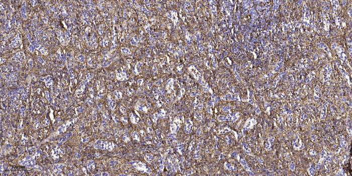
- Immunohistochemical analysis of paraffin-embedded human spleen tissue. 1,primary Antibody was diluted at 1:200(4° overnight). 2, Sodium citrate pH 6.0 was used for antigen retrieval(>98°C,20min). 3,Secondary antibody was diluted at 1:200


