IRAK-M Polyclonal Antibody
- Catalog No.:YT2393
- Applications:WB;IHC;IF;ELISA
- Reactivity:Human;Mouse
- Target:
- IRAK-M
- Fields:
- >>Neurotrophin signaling pathway
- Gene Name:
- IRAK3
- Protein Name:
- Interleukin-1 receptor-associated kinase 3
- Human Gene Id:
- 11213
- Human Swiss Prot No:
- Q9Y616
- Mouse Swiss Prot No:
- Q8K4B2
- Immunogen:
- The antiserum was produced against synthesized peptide derived from human IRAK3. AA range:491-540
- Specificity:
- IRAK-M Polyclonal Antibody detects endogenous levels of IRAK-M protein.
- Formulation:
- Liquid in PBS containing 50% glycerol, 0.5% BSA and 0.02% sodium azide.
- Source:
- Polyclonal, Rabbit,IgG
- Dilution:
- WB 1:500 - 1:2000. IHC 1:100 - 1:300. IF 1:200 - 1:1000. ELISA: 1:20000. Not yet tested in other applications.
- Purification:
- The antibody was affinity-purified from rabbit antiserum by affinity-chromatography using epitope-specific immunogen.
- Concentration:
- 1 mg/ml
- Storage Stability:
- -15°C to -25°C/1 year(Do not lower than -25°C)
- Other Name:
- IRAK3;Interleukin-1 receptor-associated kinase 3;IRAK-3;IL-1 receptor-associated kinase M;IRAK-M
- Observed Band(KD):
- 68kD
- Background:
- This gene encodes a member of the interleukin-1 receptor-associated kinase protein family. Members of this family are essential components of the Toll/IL-R immune signal transduction pathways. This protein is primarily expressed in monocytes and macrophages and functions as a negative regulator of Toll-like receptor signaling. Mutations in this gene are associated with a susceptibility to asthma. Alternate splicing results in multiple transcript variants. [provided by RefSeq, May 2010],
- Function:
- catalytic activity:ATP + a protein = ADP + a phosphoprotein.,caution:Ser-293 is present instead of the conserved Asp which is expected to be an active site residue. Low level autophosphorylation activity has been reported in PubMed:10383454, while other authors describe this as an inactive kinase.,cofactor:Magnesium.,disease:Defects in IRAK3 are associated with susceptibility to asthma-related traits type 5 (ASRT5) [MIM:611064]. Asthma-related traits include clinical symptoms of asthma, such as coughing, wheezing, dyspnea, bronchial hyperresponsiveness as assessed by methacholine challenge test, serum IgE levels, atopy, and atopic dermatitis.,function:Inhibits dissociation of IRAK1 and IRAK4 from the Toll-like receptor signaling complex by either inhibiting the phosphorylation of IRAK1 and IRAK4 or stabilizing the receptor complex.,similarity:Belongs to the protein kinase superfamily. TK
- Subcellular Location:
- Cytoplasm . Nucleus . In dendritic cells, translocates into the nucleus upon IL33 stimulation. .
- Expression:
- Expressed in eosinophils, dendritic cells and/or monocytes (at protein level) (PubMed:29686383). Expressed predominantly in peripheral blood lymphocytes (PubMed:10383454).
- June 19-2018
- WESTERN IMMUNOBLOTTING PROTOCOL
- June 19-2018
- IMMUNOHISTOCHEMISTRY-PARAFFIN PROTOCOL
- June 19-2018
- IMMUNOFLUORESCENCE PROTOCOL
- September 08-2020
- FLOW-CYTOMEYRT-PROTOCOL
- May 20-2022
- Cell-Based ELISA│解您多样本WB检测之困扰
- July 13-2018
- CELL-BASED-ELISA-PROTOCOL-FOR-ACETYL-PROTEIN
- July 13-2018
- CELL-BASED-ELISA-PROTOCOL-FOR-PHOSPHO-PROTEIN
- July 13-2018
- Antibody-FAQs
- Products Images

- Western Blot analysis of LOVO cells using IRAK-M Polyclonal Antibody

- Immunofluorescence analysis of HeLa cells, using IRAK3 Antibody. The picture on the right is blocked with the synthesized peptide.

- Immunohistochemistry analysis of paraffin-embedded human brain, using IRAK3 Antibody. The picture on the right is blocked with the synthesized peptide.
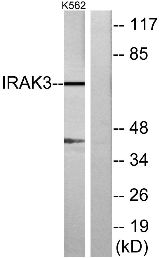
- Western blot analysis of lysates from K562 cells, using IRAK3 Antibody. The lane on the right is blocked with the synthesized peptide.

- Western blot analysis of the lysates from HUVECcells using IRAK3 antibody.


