Kpm Polyclonal Antibody
- Catalog No.:YT2494
- Applications:IHC;IF;ELISA
- Reactivity:Human;Mouse
- Target:
- Kpm
- Fields:
- >>Hippo signaling pathway;>>Hippo signaling pathway - multiple species
- Gene Name:
- LATS2
- Protein Name:
- Serine/threonine-protein kinase LATS2
- Human Gene Id:
- 26524
- Human Swiss Prot No:
- Q9NRM7
- Mouse Gene Id:
- 50523
- Mouse Swiss Prot No:
- Q7TSJ6
- Immunogen:
- The antiserum was produced against synthesized peptide derived from human LATS2. AA range:541-590
- Specificity:
- Kpm Polyclonal Antibody detects endogenous levels of Kpm protein.
- Formulation:
- Liquid in PBS containing 50% glycerol, 0.5% BSA and 0.02% sodium azide.
- Source:
- Polyclonal, Rabbit,IgG
- Dilution:
- IHC 1:100 - 1:300. IF 1:200 - 1:1000. ELISA: 1:20000. Not yet tested in other applications.
- Purification:
- The antibody was affinity-purified from rabbit antiserum by affinity-chromatography using epitope-specific immunogen.
- Concentration:
- 1 mg/ml
- Storage Stability:
- -15°C to -25°C/1 year(Do not lower than -25°C)
- Other Name:
- LATS2;KPM;Serine/threonine-protein kinase LATS2;Kinase phosphorylated during mitosis protein;Large tumor suppressor homolog 2;Serine/threonine-protein kinase kpm;Warts-like kinase
- Molecular Weight(Da):
- 120kD
- Background:
- This gene encodes a serine/threonine protein kinase belonging to the LATS tumor suppressor family. The protein localizes to centrosomes during interphase, and early and late metaphase. It interacts with the centrosomal proteins aurora-A and ajuba and is required for accumulation of gamma-tubulin and spindle formation at the onset of mitosis. It also interacts with a negative regulator of p53 and may function in a positive feedback loop with p53 that responds to cytoskeleton damage. Additionally, it can function as a co-repressor of androgen-responsive gene expression. [provided by RefSeq, Jul 2008],
- Function:
- catalytic activity:ATP + a protein = ADP + a phosphoprotein.,cofactor:Magnesium.,function:Tumor suppressor which plays a critical role in centrosome duplication, maintenance of mitotic fidelity and genomic stability. Negatively regulates G1/S transition by down-regulating cyclin E/CDK2 kinase activity. Negative regulator of the androgen receptor.,PTM:Autophosphorylated and phosphorylated during M-phase and the G1/S-phase of the cell cycle. Phosphorylated and activated by STK3.,similarity:Belongs to the protein kinase superfamily.,similarity:Belongs to the protein kinase superfamily. AGC Ser/Thr protein kinase family.,similarity:Contains 1 AGC-kinase C-terminal domain.,similarity:Contains 1 protein kinase domain.,similarity:Contains 1 UBA domain.,subcellular location:Co-localizes with STK6 at the centrosomes during interphase, early prophase and cytokinesis. Migrates to the spindle poles
- Subcellular Location:
- Cytoplasm, cytoskeleton, microtubule organizing center, centrosome. Cytoplasm. Cytoplasm, cytoskeleton, spindle pole. Nucleus. Colocalizes with AURKA at the centrosomes during interphase, early prophase and cytokinesis. Migrates to the spindle poles during mitosis, and to the midbody during cytokinesis. Translocates to the nucleus upon mitotic stress by nocodazole treatment.
- Expression:
- Expressed at high levels in heart and skeletal muscle and at lower levels in all other tissues examined.
- June 19-2018
- WESTERN IMMUNOBLOTTING PROTOCOL
- June 19-2018
- IMMUNOHISTOCHEMISTRY-PARAFFIN PROTOCOL
- June 19-2018
- IMMUNOFLUORESCENCE PROTOCOL
- September 08-2020
- FLOW-CYTOMEYRT-PROTOCOL
- May 20-2022
- Cell-Based ELISA│解您多样本WB检测之困扰
- July 13-2018
- CELL-BASED-ELISA-PROTOCOL-FOR-ACETYL-PROTEIN
- July 13-2018
- CELL-BASED-ELISA-PROTOCOL-FOR-PHOSPHO-PROTEIN
- July 13-2018
- Antibody-FAQs
- Products Images

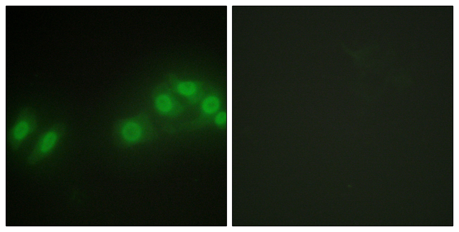
- Immunofluorescence analysis of HepG2 cells, using LATS2 Antibody. The picture on the right is blocked with the synthesized peptide.

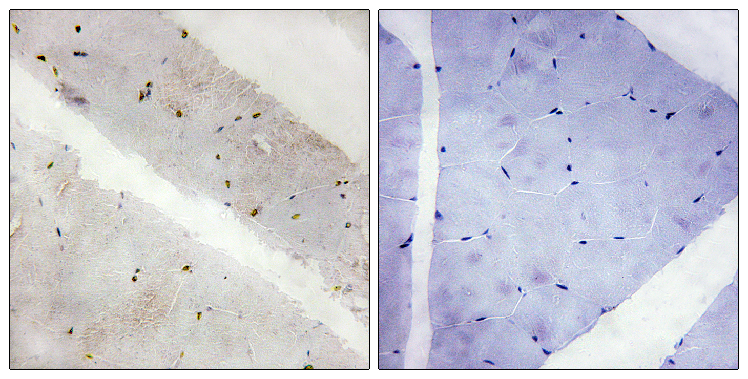
- Immunohistochemistry analysis of paraffin-embedded human skeletal muscle tissue, using LATS2 Antibody. The picture on the right is blocked with the synthesized peptide.


