Myf-6 Polyclonal Antibody
- Catalog No.:YT2931
- Applications:WB;IHC;IF;ELISA
- Reactivity:Human;Mouse;Rat
- Target:
- Myf-6
- Gene Name:
- MYF6
- Protein Name:
- Myogenic factor 6
- Human Gene Id:
- 4618
- Human Swiss Prot No:
- P23409
- Mouse Gene Id:
- 17878
- Mouse Swiss Prot No:
- P15375
- Rat Gene Id:
- 25714
- Rat Swiss Prot No:
- P19335
- Immunogen:
- The antiserum was produced against synthesized peptide derived from human MYF6. AA range:116-165
- Specificity:
- Myf-6 Polyclonal Antibody detects endogenous levels of Myf-6 protein.
- Formulation:
- Liquid in PBS containing 50% glycerol, 0.5% BSA and 0.02% sodium azide.
- Source:
- Polyclonal, Rabbit,IgG
- Dilution:
- WB 1:500 - 1:2000. IHC 1:100 - 1:300. ELISA: 1:20000.. IF 1:50-200
- Purification:
- The antibody was affinity-purified from rabbit antiserum by affinity-chromatography using epitope-specific immunogen.
- Concentration:
- 1 mg/ml
- Storage Stability:
- -15°C to -25°C/1 year(Do not lower than -25°C)
- Other Name:
- MYF6;BHLHC4;MRF4;Myogenic factor 6;Myf-6;Class C basic helix-loop-helix protein 4;bHLHc4;Muscle-specific regulatory factor 4
- Observed Band(KD):
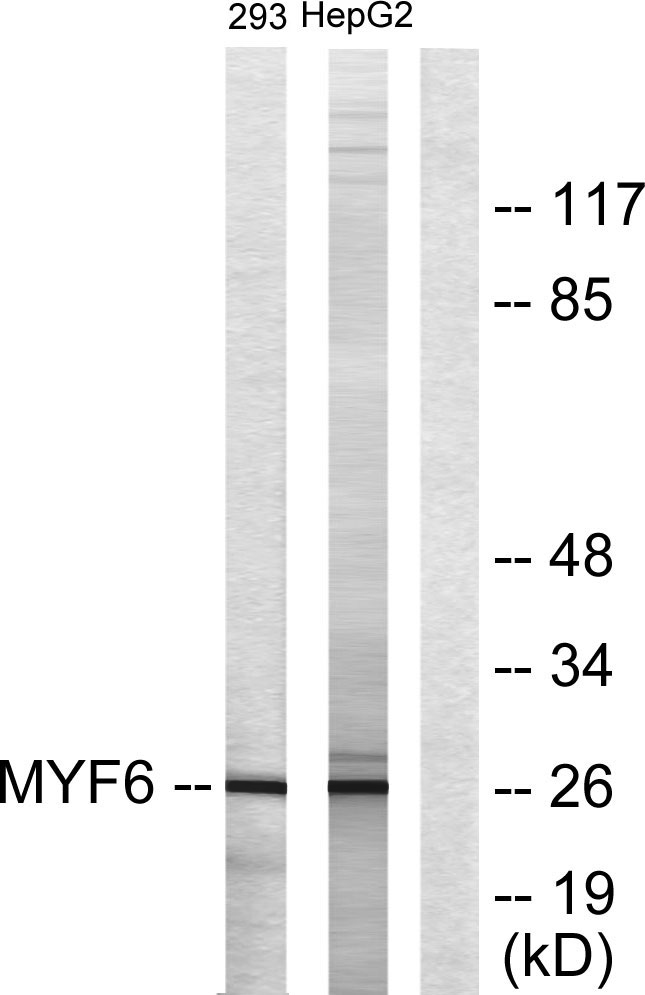
- 26kD
- Background:
- myogenic factor 6(MYF6) Homo sapiens The protein encoded by this gene is a probable basic helix-loop-helix (bHLH) DNA binding protein involved in muscle differentiation. The encoded protein likely acts as a heterodimer with another bHLH protein. Defects in this gene are a cause of autosomal dominant centronuclear myopathy (ADCNM). [provided by RefSeq, May 2010],
- Function:
- disease:Defects in MYF6 may be a cause of centronuclear myopathy autosomal dominant (ADCNM) [MIM:160150]; also known as autosomal dominant myotubular myopathy. Centronuclear myopathies are congenital muscle disorders characterized by progressive muscular weakness and wasting involving mainly limb girdle, trunk, and neck muscles. It may also affect distal muscles. Weakness may be present during childhood or adolescence or may not become evident until the third decade of life. Ptosis is a frequent clinical feature. The most prominent histopathologic features include high frequency of centrally located nuclei in muscle fibers not secondary to regeneration, radial arrangement of sarcoplasmic strands around the central nuclei, and predominance and hypotrophy of type 1 fibers.,function:Involved in muscle differentiation (myogenic factor). Induces fibroblasts to differentiate into myoblasts. Pr
- Subcellular Location:
- Nucleus.
- Expression:
- Skeletal muscle.
- June 19-2018
- WESTERN IMMUNOBLOTTING PROTOCOL
- June 19-2018
- IMMUNOHISTOCHEMISTRY-PARAFFIN PROTOCOL
- June 19-2018
- IMMUNOFLUORESCENCE PROTOCOL
- September 08-2020
- FLOW-CYTOMEYRT-PROTOCOL
- May 20-2022
- Cell-Based ELISA│解您多样本WB检测之困扰
- July 13-2018
- CELL-BASED-ELISA-PROTOCOL-FOR-ACETYL-PROTEIN
- July 13-2018
- CELL-BASED-ELISA-PROTOCOL-FOR-PHOSPHO-PROTEIN
- July 13-2018
- Antibody-FAQs
- Products Images

- Western Blot analysis of various cells using Myf-6 Polyclonal Antibody diluted at 1:2000 cells nucleus extracted by Minute TM Cytoplasmic and Nuclear Fractionation kit (SC-003,Inventbiotech,MN,USA).

- Western blot analysis of lysates from HepG2 and 293 cells, using MYF6 Antibody. The lane on the right is blocked with the synthesized peptide.

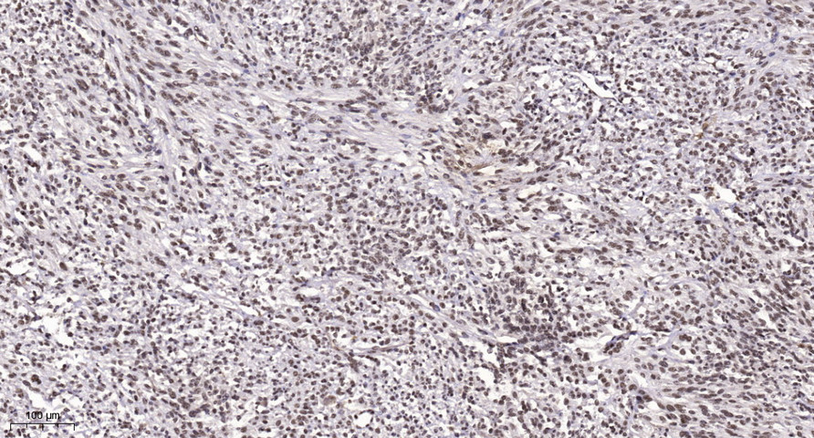
- Immunohistochemical analysis of paraffin-embedded human Small intestinal stromal tumor. 1, Tris-EDTA,pH9.0 was used for antigen retrieval. 2 Antibody was diluted at 1:200(4° overnight.3,Secondary antibody was diluted at 1:200(room temperature, 45min).


