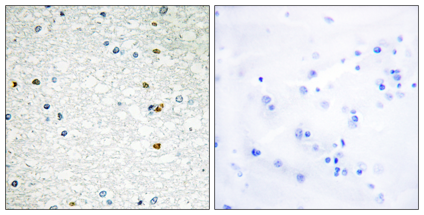
PARP-3 Polyclonal Antibody
- Catalog No.:YT3595
- Applications:IHC;IF;ELISA
- Reactivity:Human;Mouse
- Target:
- PARP-3
- Fields:
- >>Base excision repair;>>Apoptosis
- Gene Name:
- PARP3
- Protein Name:
- Poly [ADP-ribose] polymerase 3
- Human Gene Id:
- 10039
- Human Swiss Prot No:
- Q9Y6F1
- Immunogen:
- The antiserum was produced against synthesized peptide derived from human PARP3. AA range:10-59
- Specificity:
- PARP-3 Polyclonal Antibody detects endogenous levels of PARP-3 protein.
- Formulation:
- Liquid in PBS containing 50% glycerol, 0.5% BSA and 0.02% sodium azide.
- Source:
- Polyclonal, Rabbit,IgG
- Dilution:
- IHC 1:100 - 1:300. ELISA: 1:5000.. IF 1:50-200
- Purification:
- The antibody was affinity-purified from rabbit antiserum by affinity-chromatography using epitope-specific immunogen.
- Concentration:
- 1 mg/ml
- Storage Stability:
- -15°C to -25°C/1 year(Do not lower than -25°C)
- Other Name:
- PARP3;ADPRT3;ADPRTL3;Poly [ADP-ribose] polymerase 3;PARP-3;hPARP-3;ADP-ribosyltransferase diphtheria toxin-like 3;ARTD3;IRT1;NAD(+) ADP-ribosyltransferase 3;ADPRT-3;Poly[ADP-ribose] synthase 3;pADPRT-3
- Molecular Weight(Da):
- 60kD
- Background:
- The protein encoded by this gene belongs to the PARP family. These enzymes modify nuclear proteins by poly-ADP-ribosylation, which is required for DNA repair, regulation of apoptosis, and maintenance of genomic stability. This gene encodes the poly(ADP-ribosyl)transferase 3, which is preferentially localized to the daughter centriole throughout the cell cycle. Alternatively spliced transcript variants encoding different isoforms have been identified. [provided by RefSeq, Jul 2008],
- Function:
- catalytic activity:NAD(+) + (ADP-D-ribosyl)(n)-acceptor = nicotinamide + (ADP-D-ribosyl)(n+1)-acceptor.,domain:According to PubMed:10329013 the N-terminal domain (54 amino acids) of isoform 2 is responsible for its centrosomal localization. The C-terminal region contains the catalytic domain.,function:Involved in the base excision repair (BER) pathway, by catalyzing the poly(ADP-ribosyl)ation of a limited number of acceptor proteins involved in chromatin architecture and in DNA metabolism. This modification follows DNA damages and appears as an obligatory step in a detection/signaling pathway leading to the reparation of DNA strand breaks. May link the DNA damage surveillance network to the mitotic fidelity checkpoint. Negatively influences the G1/S cell cycle progression without interfering with centrosome duplication. Binds DNA. May be involved in the regulation of PRC2 and PRC3 comple
- Subcellular Location:
- Nucleus . Chromosome . Cytoplasm, cytoskeleton, microtubule organizing center, centrosome . Cytoplasm, cytoskeleton, microtubule organizing center, centrosome, centriole . Almost exclusively localized in the nucleus and appears in numerous small foci and a small number of larger foci whereas a centrosomal location has not been detected (PubMed:16924674). In response to DNA damage, localizes to sites of double-strand break (PubMed:21270334). Preferentially localized to the daughter centriole (PubMed:10329013). .
- Expression:
- Widely expressed; the highest levels are in the kidney, skeletal muscle, liver, heart and spleen; also detected in pancreas, lung, placenta, brain, leukocytes, colon, small intestine, ovary, testis, prostate and thymus.
- June 19-2018
- WESTERN IMMUNOBLOTTING PROTOCOL
- June 19-2018
- IMMUNOHISTOCHEMISTRY-PARAFFIN PROTOCOL
- June 19-2018
- IMMUNOFLUORESCENCE PROTOCOL
- September 08-2020
- FLOW-CYTOMEYRT-PROTOCOL
- May 20-2022
- Cell-Based ELISA│解您多样本WB检测之困扰
- July 13-2018
- CELL-BASED-ELISA-PROTOCOL-FOR-ACETYL-PROTEIN
- July 13-2018
- CELL-BASED-ELISA-PROTOCOL-FOR-PHOSPHO-PROTEIN
- July 13-2018
- Antibody-FAQs
- Products Images

- Immunohistochemistry analysis of paraffin-embedded human brain, using PARP3 Antibody. The picture on the right is blocked with the synthesized peptide.


