PDPK1 Polyclonal Antibody
- Catalog No.:YT3645
- Applications:IF;WB;IHC;ELISA
- Reactivity:Human;Mouse;Rat
- Target:
- PDPK1
- Fields:
- >>Platinum drug resistance;>>PPAR signaling pathway;>>FoxO signaling pathway;>>Sphingolipid signaling pathway;>>Autophagy - animal;>>mTOR signaling pathway;>>PI3K-Akt signaling pathway;>>AMPK signaling pathway;>>Apoptosis;>>Axon guidance;>>Focal adhesion;>>T cell receptor signaling pathway;>>Fc epsilon RI signaling pathway;>>Neurotrophin signaling pathway;>>Insulin signaling pathway;>>Thyroid hormone signaling pathway;>>Insulin resistance;>>Aldosterone-regulated sodium reabsorption;>>Toxoplasmosis;>>Proteoglycans in cancer;>>Chemical carcinogenesis - reactive oxygen species;>>Endometrial cancer;>>Prostate cancer;>>Non-small cell lung cancer;>>Choline metabolism in cancer;>>Lipid and atherosclerosis
- Gene Name:
- PDPK1
- Protein Name:
- 3-phosphoinositide-dependent protein kinase 1
- Human Gene Id:
- 5170
- Human Swiss Prot No:
- O15530
- Mouse Gene Id:
- 18607
- Mouse Swiss Prot No:
- Q9Z2A0
- Rat Gene Id:
- 81745
- Rat Swiss Prot No:
- O55173
- Immunogen:
- The antiserum was produced against synthesized peptide derived from human PDK1. AA range:210-259
- Specificity:
- PDK1 Polyclonal Antibody detects endogenous levels of PDK1 protein.
- Formulation:
- Liquid in PBS containing 50% glycerol, 0.5% BSA and 0.02% sodium azide.
- Source:
- Polyclonal, Rabbit,IgG
- Dilution:
- IF 1:50-200 WB 1:500 - 1:2000. IHC 1:100 - 1:300. ELISA: 1:10000. Not yet tested in other applications.
- Purification:
- The antibody was affinity-purified from rabbit antiserum by affinity-chromatography using epitope-specific immunogen.
- Concentration:
- 1 mg/ml
- Storage Stability:
- -15°C to -25°C/1 year(Do not lower than -25°C)
- Other Name:
- PDPK1;PDK1;3-phosphoinositide-dependent protein kinase 1;hPDK1
- Observed Band(KD):
- 60kD
- Background:
- catalytic activity:ATP + a protein = ADP + a phosphoprotein.,function:Phosphorylates and activates not only PKB/AKT, but also PKA, PKC-zeta, RPS6KA1 and RPS6KB1. May play a general role in signaling processes and in development (By similarity). Isoform 3 is catalytically inactive.,PTM:Phosphorylated on tyrosine and serine/threonine. Phosphorylation on Ser-241 in the activation loop is required for full activity. PDK1 itself can autophosphorylate Ser-241, leading to its own activation.,similarity:Belongs to the protein kinase superfamily.,similarity:Belongs to the protein kinase superfamily. AGC Ser/Thr protein kinase family. PDK1 subfamily.,similarity:Contains 1 PH domain.,similarity:Contains 1 protein kinase domain.,subcellular location:Membrane-associated after cell stimulation leading to its translocation. Tyrosine phosphorylation seems to occur only at the plasma membrane.,subunit:Interacts with TUSC4.,tissue specificity:Appears to be expressed ubiquitously.,
- Function:
- catalytic activity:ATP + a protein = ADP + a phosphoprotein.,function:Phosphorylates and activates not only PKB/AKT, but also PKA, PKC-zeta, RPS6KA1 and RPS6KB1. May play a general role in signaling processes and in development (By similarity). Isoform 3 is catalytically inactive.,PTM:Phosphorylated on tyrosine and serine/threonine. Phosphorylation on Ser-241 in the activation loop is required for full activity. PDK1 itself can autophosphorylate Ser-241, leading to its own activation.,similarity:Belongs to the protein kinase superfamily.,similarity:Belongs to the protein kinase superfamily. AGC Ser/Thr protein kinase family. PDK1 subfamily.,similarity:Contains 1 PH domain.,similarity:Contains 1 protein kinase domain.,subcellular location:Membrane-associated after cell stimulation leading to its translocation. Tyrosine phosphorylation seems to occur only at the plasma membrane.,subunit:In
- Subcellular Location:
- Cytoplasm. Nucleus. Cell membrane; Peripheral membrane protein. Cell junction, focal adhesion. Tyrosine phosphorylation seems to occur only at the cell membrane. Translocates to the cell membrane following insulin stimulation by a mechanism that involves binding to GRB14 and INSR. SRC and HSP90 promote its localization to the cell membrane. Its nuclear localization is dependent on its association with PTPN6 and its phosphorylation at Ser-396. Restricted to the nucleus in neuronal cells while in non-neuronal cells it is found in the cytoplasm. The Ser-241 phosphorylated form is distributed along the perinuclear region in neuronal cells while in non-neuronal cells it is found in both the nucleus and the cytoplasm. IGF1 transiently increases phosphorylation at Ser-241 of neuronal PDPK1, resul
- Expression:
- Appears to be expressed ubiquitously. The Tyr-9 phosphorylated form is markedly increased in diseased tissue compared with normal tissue from lung, liver, colon and breast.
Melatonin treatment improves human umbilical cord mesenchymal stem cell therapy in a mouse model of type II diabetes mellitus via the PI3K/AKT signaling pathway. Stem Cell Research & Therapy2022 Dec;13(1):1-15. Human,Mouse 1:1200 liver tissue hUC-MSC
CD38 is involved in cell energy metabolism via activating the PI3K/AKT/mTOR signaling pathway in cervical cancer cells. INTERNATIONAL JOURNAL OF ONCOLOGY Int J Oncol. 2020 Jul;57(1):338-354 WB Human 1:1000 CaSki cell,Hela cell
CD90 affects the biological behavior and energy metabolism level of gastric cancer cells by targeting the PI3K/AKT/HIF‑1α signaling pathway. Oncology Letters Oncol Lett. 2021 Mar;21(3):1-1 WB Human 1:1000 AGS cell
MAOA suppresses the growth of gastric cancer by interacting with NDRG1 and regulating the Warburg effect through the PI3K/AKT/mTOR pathway. Li Jun WB Human 1:1000 BGC-823 cell,HGC-27 cell
- June 19-2018
- WESTERN IMMUNOBLOTTING PROTOCOL
- June 19-2018
- IMMUNOHISTOCHEMISTRY-PARAFFIN PROTOCOL
- June 19-2018
- IMMUNOFLUORESCENCE PROTOCOL
- September 08-2020
- FLOW-CYTOMEYRT-PROTOCOL
- May 20-2022
- Cell-Based ELISA│解您多样本WB检测之困扰
- July 13-2018
- CELL-BASED-ELISA-PROTOCOL-FOR-ACETYL-PROTEIN
- July 13-2018
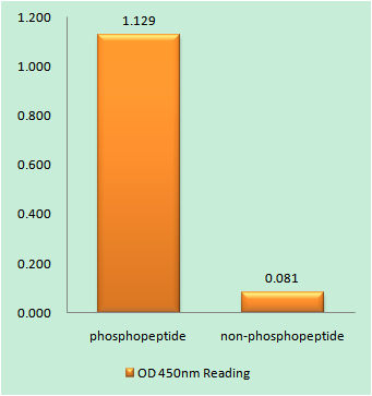
- CELL-BASED-ELISA-PROTOCOL-FOR-PHOSPHO-PROTEIN
- July 13-2018
- Antibody-FAQs
- Products Images

- Western blot analysis of lysates from 1) Hela, 2) COLO,3) 293T cells, (Green) primary antibody was diluted at 1:1000, 4°over night, secondary antibody(cat:RS23920)was diluted at 1:10000, 37° 1hour. (Red) GAPDH Monoclonal Antibody(2B8) (cat:YM3029) antibody was diluted at 1:5000 as loading control, 4° over night,secondary antibody(cat:RS23710)was diluted at 1:10000, 37° 1hour.
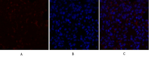
- Immunofluorescence analysis of rat-lung tissue. 1,PDK1 Polyclonal Antibody(red) was diluted at 1:200(4°C,overnight). 2, Cy3 labled Secondary antibody was diluted at 1:300(room temperature, 50min).3, Picture B: DAPI(blue) 10min. Picture A:Target. Picture B: DAPI. Picture C: merge of A+B
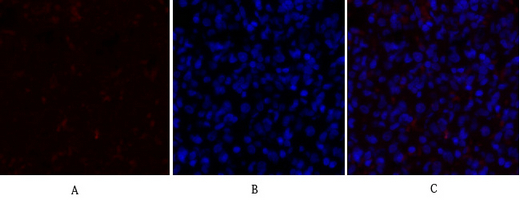
- Immunofluorescence analysis of rat-lung tissue. 1,PDK1 Polyclonal Antibody(red) was diluted at 1:200(4°C,overnight). 2, Cy3 labled Secondary antibody was diluted at 1:300(room temperature, 50min).3, Picture B: DAPI(blue) 10min. Picture A:Target. Picture B: DAPI. Picture C: merge of A+B
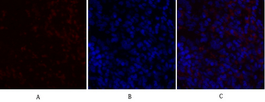
- Immunofluorescence analysis of rat-spleen tissue. 1,PDK1 Polyclonal Antibody(red) was diluted at 1:200(4°C,overnight). 2, Cy3 labled Secondary antibody was diluted at 1:300(room temperature, 50min).3, Picture B: DAPI(blue) 10min. Picture A:Target. Picture B: DAPI. Picture C: merge of A+B

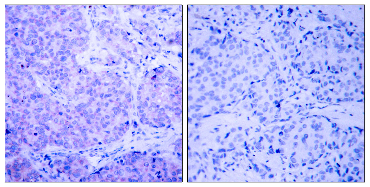
- Immunohistochemistry analysis of paraffin-embedded human breast carcinoma tissue, using PDK1 Antibody. The picture on the right is blocked with the synthesized peptide.
