PTG Polyclonal Antibody
- Catalog No.:YT3895
- Applications:WB;IHC;IF;ELISA
- Reactivity:Human;Monkey
- Target:
- PTG
- Fields:
- >>Insulin signaling pathway;>>Insulin resistance
- Gene Name:
- PPP1R3C
- Protein Name:
- Protein phosphatase 1 regulatory subunit 3C
- Human Gene Id:
- 5507
- Human Swiss Prot No:
- Q9UQK1
- Mouse Swiss Prot No:
- Q7TMB3
- Immunogen:
- The antiserum was produced against synthesized peptide derived from human PPP1R3C. AA range:44-93
- Specificity:
- PTG Polyclonal Antibody detects endogenous levels of PTG protein.
- Formulation:
- Liquid in PBS containing 50% glycerol, 0.5% BSA and 0.02% sodium azide.
- Source:
- Polyclonal, Rabbit,IgG
- Dilution:
- WB 1:500 - 1:2000. IHC 1:100 - 1:300. ELISA: 1:20000.. IF 1:50-200
- Purification:
- The antibody was affinity-purified from rabbit antiserum by affinity-chromatography using epitope-specific immunogen.
- Concentration:
- 1 mg/ml
- Storage Stability:
- -15°C to -25°C/1 year(Do not lower than -25°C)
- Other Name:
- PPP1R3C;PPP1R5;Protein phosphatase 1 regulatory subunit 3C;Protein phosphatase 1 regulatory subunit 5;PP1 subunit R5;Protein targeting to glycogen;PTG
- Observed Band(KD):
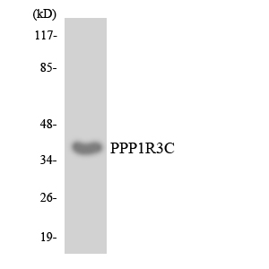
- 36kD
- Background:
- This gene encodes a carbohydrate binding protein that is a subunit of the protein phosphatase 1 (PP1) complex. PP1 catalyzes reversible protein phosphorylation, which is important in a wide range of cellular activities. The encoded protein affects glycogen biosynthesis by activating glycogen synthase and limiting glycogen breakdown by reducing glycogen phosphorylase activity. DNA hypermethylation of this gene has been found in colorectal cancer patients. The encoded protein also interacts with the laforin protein, which is a protein tyrosine phosphatase implicated in Lafora disease. [provided by RefSeq, Sep 2016],
- Function:
- domain:The N-terminal region is required for binding to PP1, the central region is required for binding to glycogen and the C-terminal region is required for binding to glycogen phosphorylase, glycogen synthase and phosphorylase kinase.,function:Acts as a glycogen-targeting subunit for PP1 and regulates its activity. Activates glycogen synthase, reduces glycogen phosphorylase activity and limits glycogen breakdown. Dramatically increases basal and insulin-stimulated glycogen synthesis upon overexpression in a variety of cell types.,similarity:Contains 1 CBM21 (carbohydrate binding type-21) domain.,subunit:Interacts with PPP1CC catalytic subunit of PP1 and associates with glycogen. Forms complexes with glycogen phosphorylase, glycogen synthase and phosphorylase kinase which is necessary for its regulation of PP1 activity. Also interacts with EPM2A/laforin.,
- Subcellular Location:
- cytosol,
- Expression:
- Gall bladder,Retina,Skeletal muscle,
- June 19-2018
- WESTERN IMMUNOBLOTTING PROTOCOL
- June 19-2018
- IMMUNOHISTOCHEMISTRY-PARAFFIN PROTOCOL
- June 19-2018
- IMMUNOFLUORESCENCE PROTOCOL
- September 08-2020
- FLOW-CYTOMEYRT-PROTOCOL
- May 20-2022
- Cell-Based ELISA│解您多样本WB检测之困扰
- July 13-2018
- CELL-BASED-ELISA-PROTOCOL-FOR-ACETYL-PROTEIN
- July 13-2018
- CELL-BASED-ELISA-PROTOCOL-FOR-PHOSPHO-PROTEIN
- July 13-2018
- Antibody-FAQs
- Products Images

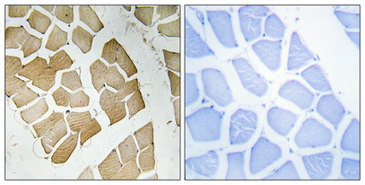
- Immunohistochemical analysis of paraffin-embedded Human skeletal muscle. Antibody was diluted at 1:100(4° overnight). High-pressure and temperature Tris-EDTA,pH8.0 was used for antigen retrieval. Negetive contrl (right) obtaned from antibody was pre-absorbed by immunogen peptide.

- Western blot analysis of lysates from HepG2, HeLa, and COS7 cells, using PPP1R3C Antibody. The lane on the right is blocked with the synthesized peptide.

- Western blot analysis of the lysates from HeLa cells using PPP1R3C antibody.


