Rad17 Polyclonal Antibody
- Catalog No.:YT3957
- Applications:WB;IHC;IF;ELISA
- Reactivity:Human;Mouse
- Target:
- Rad17
- Gene Name:
- RAD17
- Protein Name:
- Cell cycle checkpoint protein RAD17
- Human Gene Id:
- 5884
- Human Swiss Prot No:
- O75943
- Mouse Gene Id:
- 19356
- Mouse Swiss Prot No:
- Q6NXW6
- Immunogen:
- The antiserum was produced against synthesized peptide derived from human RAD17. AA range:621-670
- Specificity:
- Rad17 Polyclonal Antibody detects endogenous levels of Rad17 protein.
- Formulation:
- Liquid in PBS containing 50% glycerol, 0.5% BSA and 0.02% sodium azide.
- Source:
- Polyclonal, Rabbit,IgG
- Dilution:
- WB 1:500 - 1:2000. IHC 1:100 - 1:300. IF 1:200 - 1:1000. ELISA: 1:10000. Not yet tested in other applications.
- Purification:
- The antibody was affinity-purified from rabbit antiserum by affinity-chromatography using epitope-specific immunogen.
- Concentration:
- 1 mg/ml
- Storage Stability:
- -15°C to -25°C/1 year(Do not lower than -25°C)
- Other Name:
- RAD17;R24L;Cell cycle checkpoint protein RAD17;hRad17;RF-C/activator 1 homolog
- Observed Band(KD):
- 86kD
- Background:
- The protein encoded by this gene is highly similar to the gene product of Schizosaccharomyces pombe rad17, a cell cycle checkpoint gene required for cell cycle arrest and DNA damage repair in response to DNA damage. This protein shares strong similarity with DNA replication factor C (RFC), and can form a complex with RFCs. This protein binds to chromatin prior to DNA damage and is phosphorylated by the checkpoint kinase ATR following damage. This protein recruits the RAD1-RAD9-HUS1 checkpoint protein complex onto chromatin after DNA damage, which may be required for its phosphorylation. The phosphorylation of this protein is required for the DNA-damage-induced cell cycle G2 arrest, and is thought to be a critical early event during checkpoint signaling in DNA-damaged cells. Multiple alternatively spliced transcript variants of this gene, which encode four distinct protein isoforms, h
- Function:
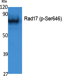
- function:Essential for sustained cell growth, maintenance of chromosomal stability, and ATR-dependent checkpoint activation upon DNA damage. Has a weak ATPase activity required for binding to chromatin. Participates in the recruitment of the RAD1-RAD9-HUS1 complex onto chromatin, and in CHEK1 activation. May also serve as a sensor of DNA replication progression, and may be involved in homologous recombination.,induction:By X-ray irradiation (isoform 1, isoform 3 and isoform 4).,PTM:Phosphorylated. Phosphorylation on Ser-646 and Ser-656 is cell cycle-regulated, enhanced by genotoxic stress, and required for activation of checkpoint signaling. Phosphorylation is mediated by ATR upon UV or replication arrest, whereas it may be mediated both by ATR and ATM upon ionizing radiation. Phosphorylation on both sites is required for interaction with RAD1 but dispensable for interaction with RFC3 or
- Subcellular Location:
- Nucleus . Phosphorylated form redistributes to discrete nuclear foci upon DNA damage.
- Expression:
- Overexpressed in various cancer cell lines and in colon carcinoma (at protein level). Isoform 2 and isoform 3 are the most abundant isoforms in non irradiated cells (at protein level). Ubiquitous at low levels. Highly expressed in testis, where it is expressed within the germinal epithelium of the seminiferous tubuli. Weakly expressed in seminomas (testicular tumors).
- June 19-2018
- WESTERN IMMUNOBLOTTING PROTOCOL
- June 19-2018
- IMMUNOHISTOCHEMISTRY-PARAFFIN PROTOCOL
- June 19-2018
- IMMUNOFLUORESCENCE PROTOCOL
- September 08-2020
- FLOW-CYTOMEYRT-PROTOCOL
- May 20-2022
- Cell-Based ELISA│解您多样本WB检测之困扰
- July 13-2018
- CELL-BASED-ELISA-PROTOCOL-FOR-ACETYL-PROTEIN
- July 13-2018
- CELL-BASED-ELISA-PROTOCOL-FOR-PHOSPHO-PROTEIN
- July 13-2018
- Antibody-FAQs
- Products Images
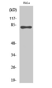
- Western Blot analysis of various cells using Rad17 Polyclonal Antibody
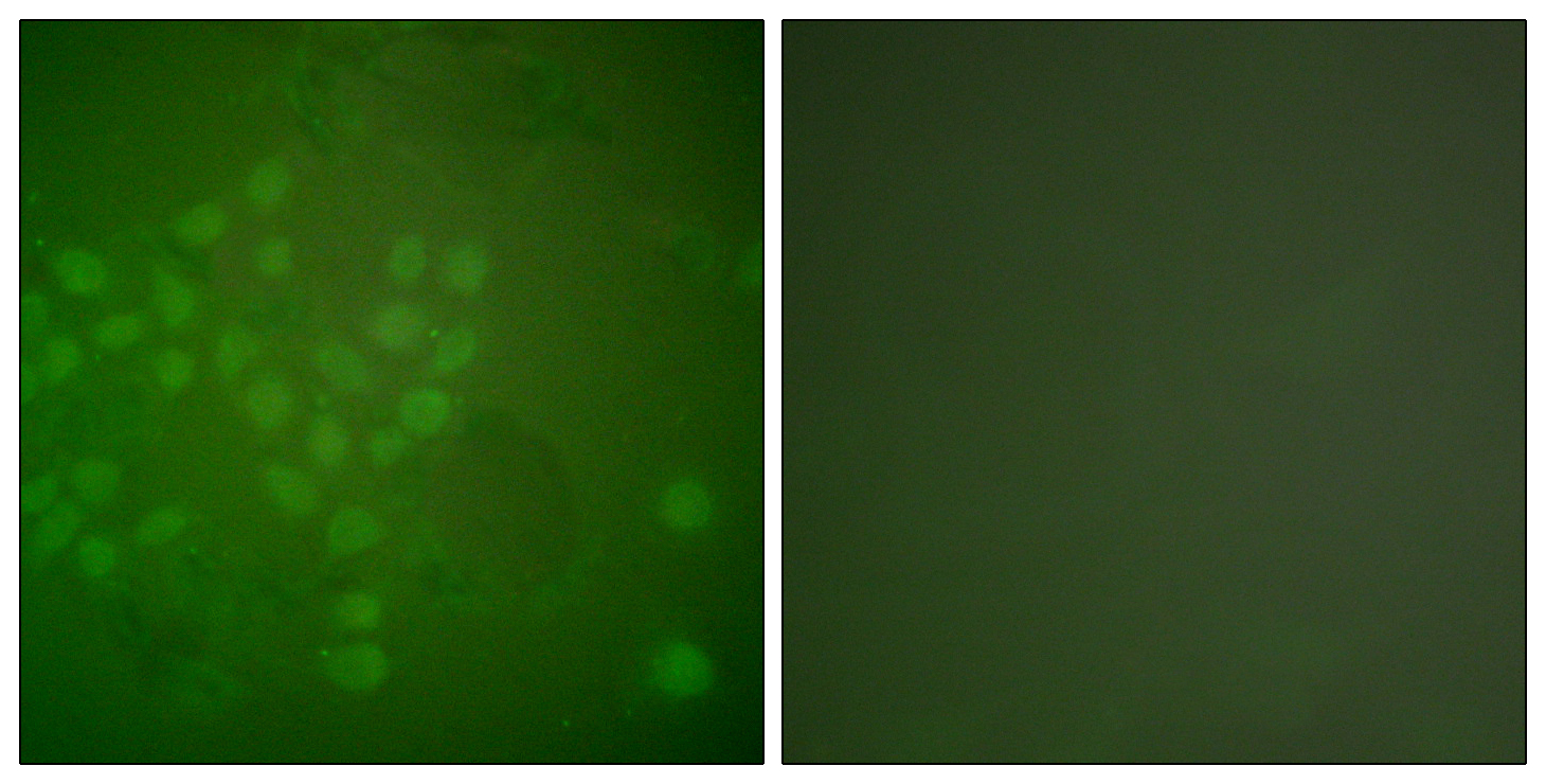
- Immunofluorescence analysis of A549 cells, using RAD17 Antibody. The picture on the right is blocked with the synthesized peptide.
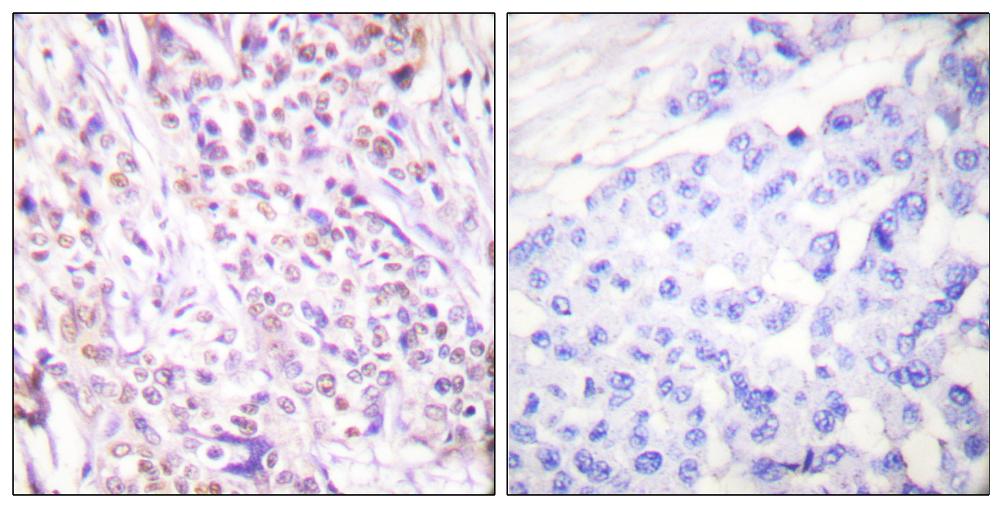
- Immunohistochemistry analysis of paraffin-embedded human breast carcinoma tissue, using RAD17 Antibody. The picture on the right is blocked with the synthesized peptide.
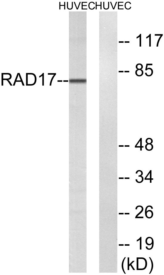
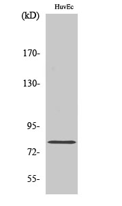
- Western blot analysis of lysates from HUVEC cells, using RAD17 Antibody. The lane on the right is blocked with the synthesized peptide.