Rad51C Polyclonal Antibody
- Catalog No.:YT3967
- Applications:WB;IHC;IF;ELISA
- Reactivity:Human;Monkey
- Target:
- Rad51C
- Fields:
- >>Homologous recombination;>>Fanconi anemia pathway
- Gene Name:
- RAD51C
- Protein Name:
- DNA repair protein RAD51 homolog 3
- Human Gene Id:
- 5889
- Human Swiss Prot No:
- O43502
- Mouse Swiss Prot No:
- Q924H5
- Immunogen:
- The antiserum was produced against synthesized peptide derived from human RAD51C. AA range:161-210
- Specificity:
- Rad51C Polyclonal Antibody detects endogenous levels of Rad51C protein.
- Formulation:
- Liquid in PBS containing 50% glycerol, 0.5% BSA and 0.02% sodium azide.
- Source:
- Polyclonal, Rabbit,IgG
- Dilution:
- WB 1:500 - 1:2000. IHC 1:100 - 1:300. IF 1:200 - 1:1000. ELISA: 1:5000. Not yet tested in other applications.
- Purification:
- The antibody was affinity-purified from rabbit antiserum by affinity-chromatography using epitope-specific immunogen.
- Concentration:
- 1 mg/ml
- Storage Stability:
- -15°C to -25°C/1 year(Do not lower than -25°C)
- Other Name:
- RAD51C;RAD51L2;DNA repair protein RAD51 homolog 3;R51H3;RAD51 homolog C;RAD51-like protein 2
- Observed Band(KD):
- 50kD
- Background:
- RAD51 paralog C(RAD51C) Homo sapiens This gene is a member of the RAD51 family. RAD51 family members are highly similar to bacterial RecA and Saccharomyces cerevisiae Rad51 and are known to be involved in the homologous recombination and repair of DNA. This protein can interact with other RAD51 paralogs and is reported to be important for Holliday junction resolution. Mutations in this gene are associated with Fanconi anemia-like syndrome. This gene is one of four localized to a region of chromosome 17q23 where amplification occurs frequently in breast tumors. Overexpression of the four genes during amplification has been observed and suggests a possible role in tumor progression. Alternative splicing results in multiple transcript variants. [provided by RefSeq, Jul 2013],
- Function:
- function:Involved in the homologous recombination repair (HRR) pathway of double-stranded DNA breaks arising during DNA replication or induced by DNA-damaging agents. The RAD51B-RAD51C dimer exhibits single-stranded DNA-dependent ATPase activity. The BCDX2 complex binds single-stranded DNA, single-stranded gaps in duplex DNA and specifically to nicks in duplex DNA.,similarity:Belongs to the recA family. RAD51 subfamily.,subunit:Interacts with RAD51B and XRCC3. Part of a BCDX2 complex consisting of RAD51B, RAD51C, RAD51D and XRCC2. Part of a complex consisting of RAD51B, RAD51C, RAD51D, XRCC2 and XRCC3. Part of a complex with RAD51B and RAD51.,tissue specificity:Expressed in a variety of tissues, with highest expression in testis, heart muscle, spleen and prostate.,
- Subcellular Location:
- Nucleus . Cytoplasm . Cytoplasm, perinuclear region . Mitochondrion . DNA damage induces an increase in nuclear levels. Accumulates in DNA damage induced nuclear foci or RAD51C foci which is formed during the S or G2 phase of cell cycle. Accumulation at DNA lesions requires the presence of NBN/NBS1, ATM and RPA.
- Expression:
- Expressed in a variety of tissues, with highest expression in testis, heart muscle, spleen and prostate.
Antitumor effects and mechanisms of olaparib in combination with carboplatin and BKM120 on human triple‑negative breast cancer cells. ONCOLOGY REPORTS 2018 Sep 20 IF Human CAL51cell
- June 19-2018
- WESTERN IMMUNOBLOTTING PROTOCOL
- June 19-2018
- IMMUNOHISTOCHEMISTRY-PARAFFIN PROTOCOL
- June 19-2018
- IMMUNOFLUORESCENCE PROTOCOL
- September 08-2020
- FLOW-CYTOMEYRT-PROTOCOL
- May 20-2022
- Cell-Based ELISA│解您多样本WB检测之困扰
- July 13-2018
- CELL-BASED-ELISA-PROTOCOL-FOR-ACETYL-PROTEIN
- July 13-2018
- CELL-BASED-ELISA-PROTOCOL-FOR-PHOSPHO-PROTEIN
- July 13-2018
- Antibody-FAQs
- Products Images

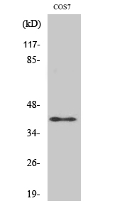
- Western Blot analysis of various cells using Rad51C Polyclonal Antibody cells nucleus extracted by Minute TM Cytoplasmic and Nuclear Fractionation kit (SC-003,Inventbiotech,MN,USA).

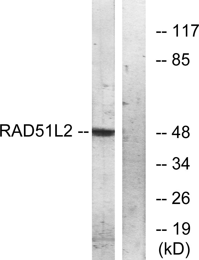
- Western blot analysis of lysates from COS7 cells, using RAD51L2 Antibody. The lane on the right is blocked with the synthesized peptide.

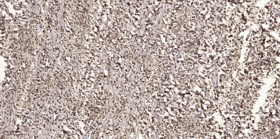
- Immunohistochemical analysis of paraffin-embedded human small intestinal carcinoma tissue. 1,primary Antibody was diluted at 1:200(4° overnight). 2, Sodium citrate pH 6.0 was used for antigen retrieval(>98°C,20min). 3,Secondary antibody was diluted at 1:200


