Ribosomal Protein S6 Polyclonal Antibody
- Catalog No.:YT4139
- Applications:WB;IHC;IF;ELISA
- Reactivity:Human;Mouse;Rat
- Target:
- Ribosomal Protein S6
- Fields:
- >>EGFR tyrosine kinase inhibitor resistance;>>Ribosome;>>HIF-1 signaling pathway;>>mTOR signaling pathway;>>PI3K-Akt signaling pathway;>>Apelin signaling pathway;>>Thermogenesis;>>Insulin signaling pathway;>>Coronavirus disease - COVID-19;>>Proteoglycans in cancer
- Gene Name:
- RPS6
- Protein Name:
- 40S ribosomal protein S6
- Human Gene Id:
- 6194
- Human Swiss Prot No:
- P62753
- Mouse Gene Id:
- 20104
- Mouse Swiss Prot No:
- P62754
- Rat Gene Id:
- 1.00911e+008
- Rat Swiss Prot No:
- P62755
- Immunogen:
- The antiserum was produced against synthesized peptide derived from human S6 Ribosomal Protein. AA range:191-240
- Specificity:
- Ribosomal Protein S6 Polyclonal Antibody detects endogenous levels of Ribosomal Protein S6 protein.
- Formulation:
- Liquid in PBS containing 50% glycerol, 0.5% BSA and 0.02% sodium azide.
- Source:
- Polyclonal, Rabbit,IgG
- Dilution:
- WB 1:500 - 1:2000. IHC 1:100 - 1:300. IF 1:200 - 1:1000. ELISA: 1:5000. Not yet tested in other applications.
- Purification:
- The antibody was affinity-purified from rabbit antiserum by affinity-chromatography using epitope-specific immunogen.
- Concentration:
- 1 mg/ml
- Storage Stability:
- -15°C to -25°C/1 year(Do not lower than -25°C)
- Other Name:
- RPS6;OK/SW-cl.2;40S ribosomal protein S6;Phosphoprotein NP33
- Observed Band(KD):
- 28kD
- Background:
- Ribosomes, the organelles that catalyze protein synthesis, consist of a small 40S subunit and a large 60S subunit. Together these subunits are composed of 4 RNA species and approximately 80 structurally distinct proteins. This gene encodes a cytoplasmic ribosomal protein that is a component of the 40S subunit. The protein belongs to the S6E family of ribosomal proteins. It is the major substrate of protein kinases in the ribosome, with subsets of five C-terminal serine residues phosphorylated by different protein kinases. Phosphorylation is induced by a wide range of stimuli, including growth factors, tumor-promoting agents, and mitogens. Dephosphorylation occurs at growth arrest. The protein may contribute to the control of cell growth and proliferation through the selective translation of particular classes of mRNA. As is typical for genes encoding ribosomal proteins, there are multiple processed
- Function:
- function:May play an important role in controlling cell growth and proliferation through the selective translation of particular classes of mRNA.,PTM:Ribosomal protein S6 is the major substrate of protein kinases in eukaryote ribosomes. The phosphorylation is stimulated by growth factors, tumor promoting agents, and mitogens. It is dephosphorylated at growth arrest.,similarity:Belongs to the ribosomal protein S6e family.,
- Subcellular Location:
- nucleus,nucleoplasm,nucleolus,cytoplasm,cytosol,ribosome,polysome,small ribosomal subunit,membrane,cytosolic small ribosomal subunit,dendrite,intracellular ribonucleoprotein complex,cytoplasmic ribonucleoprotein granu
- Expression:
- Brain,Colon,Colon adenocarcinoma,Epithelium,Muscle,Ovary,Pancreas,Placenta,Skin,Tes
Lipotoxicity-induced STING1 activation stimulates MTORC1 and restricts hepatic lipophagy. Autophagy 2021 Aug 12 WB Human Hep3B cells
Peroxisome proliferator‑activated receptor‑γ agonist inhibits the mammalian target of rapamycin signaling pathway and has a protective effect in a rat model of status epilepticus. Molecular Medicine Reports Mol Med Rep. 2015 Aug;12(2):1877-1883 WB Rat 1:500 hippocampal tissues
- June 19-2018
- WESTERN IMMUNOBLOTTING PROTOCOL
- June 19-2018
- IMMUNOHISTOCHEMISTRY-PARAFFIN PROTOCOL
- June 19-2018
- IMMUNOFLUORESCENCE PROTOCOL
- September 08-2020
- FLOW-CYTOMEYRT-PROTOCOL
- May 20-2022
- Cell-Based ELISA│解您多样本WB检测之困扰
- July 13-2018
- CELL-BASED-ELISA-PROTOCOL-FOR-ACETYL-PROTEIN
- July 13-2018
- CELL-BASED-ELISA-PROTOCOL-FOR-PHOSPHO-PROTEIN
- July 13-2018
- Antibody-FAQs
- Products Images
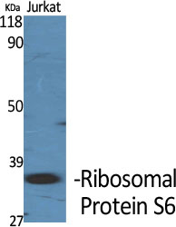
- Western Blot analysis of various cells using Ribosomal Protein S6 Polyclonal Antibody diluted at 1:2000
.jpg)
- Western Blot analysis of HeLa cells using Ribosomal Protein S6 Polyclonal Antibody diluted at 1:2000
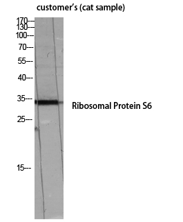
- Western Blot analysis of customer's (cat sample) using Ribosomal Protein S6 Polyclonal Antibody diluted at 1:2000
- The picture was kindly provided by our customer

- Immunofluorescence analysis of HeLa cells, using S6 Ribosomal Protein Antibody. The picture on the right is blocked with the synthesized peptide.
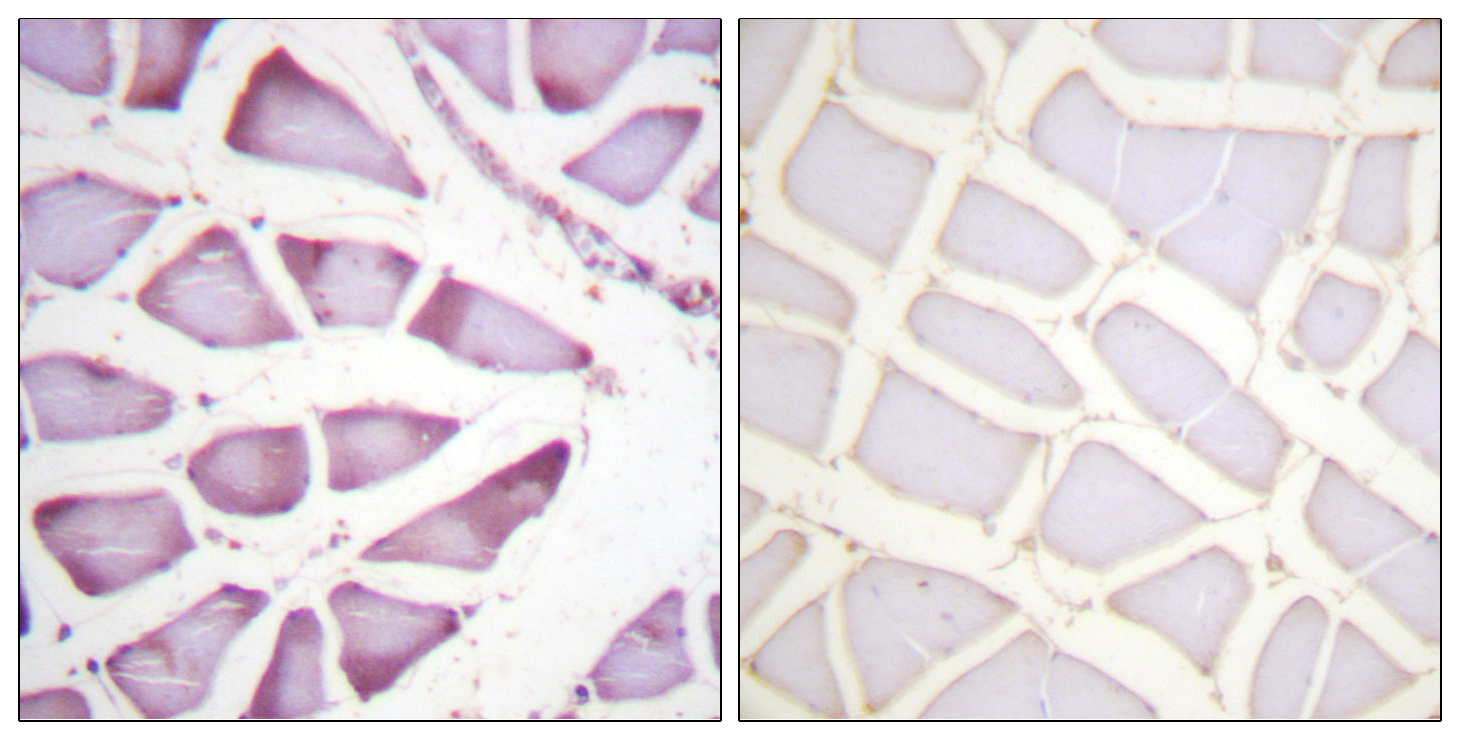
- Immunohistochemistry analysis of paraffin-embedded human skeletal muscle tissue, using S6 Ribosomal Protein Antibody. The picture on the right is blocked with the synthesized peptide.

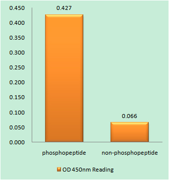
- Western blot analysis of lysates from HeLa cells, treated with TNF-a 20ng/ml 2', using S6 Ribosomal Protein Antibody. The lane on the right is blocked with the synthesized peptide.