TPX2 Polyclonal Antibody
- Catalog No.:YT4712
- Applications:WB;IHC;IF;ELISA
- Reactivity:Human;Mouse
- Target:
- TPX2
- Gene Name:
- TPX2
- Protein Name:
- Targeting protein for Xklp2
- Human Gene Id:
- 22974
- Human Swiss Prot No:
- Q9ULW0
- Mouse Gene Id:
- 72119
- Mouse Swiss Prot No:
- A2APB8
- Immunogen:
- The antiserum was produced against synthesized peptide derived from human DIL-2. AA range:301-350
- Specificity:
- TPX2 Polyclonal Antibody detects endogenous levels of TPX2 protein.
- Formulation:
- Liquid in PBS containing 50% glycerol, 0.5% BSA and 0.02% sodium azide.
- Source:
- Polyclonal, Rabbit,IgG
- Dilution:
- WB 1:500 - 1:2000. IHC 1:100 - 1:300. ELISA: 1:10000.. IF 1:50-200
- Purification:
- The antibody was affinity-purified from rabbit antiserum by affinity-chromatography using epitope-specific immunogen.
- Concentration:
- 1 mg/ml
- Storage Stability:
- -15°C to -25°C/1 year(Do not lower than -25°C)
- Other Name:
- TPX2;C20orf1;C20orf2;DIL2;HCA519;Targeting protein for Xklp2;Differentially expressed in cancerous and non-cancerous lung cells 2;DIL-2;Hepatocellular carcinoma-associated antigen 519;Protein fls353;Restricted expression prolifera
- Observed Band(KD):
- 86kD
- Background:
- developmental stage:Exclusively expressed in proliferating cells from the transition G1/S until the end of cytokinesis.,PTM:Phosphorylated upon DNA damage, probably by ATM or ATR.,subcellular location:During mitosis it is strictly associated with the spindle pole and with the mitotic spindle, whereas during S and G2, it is diffusely distributed throughout the nucleus.,tissue specificity:Expressed in lung carcinoma cell lines but not in normal lung tissues.,
- Function:
- developmental stage:Exclusively expressed in proliferating cells from the transition G1/S until the end of cytokinesis.,PTM:Phosphorylated upon DNA damage, probably by ATM or ATR.,subcellular location:During mitosis it is strictly associated with the spindle pole and with the mitotic spindle, whereas during S and G2, it is diffusely distributed throughout the nucleus.,tissue specificity:Expressed in lung carcinoma cell lines but not in normal lung tissues.,
- Subcellular Location:
- Nucleus . Cytoplasm, cytoskeleton, spindle . Cytoplasm, cytoskeleton, spindle pole . During mitosis it is strictly associated with the spindle pole and with the mitotic spindle, whereas during S and G2, it is diffusely distributed throughout the nucleus. Is released from the nucleus in apoptotic cells and is detected on apoptotic microtubules. .
- Expression:
- Expressed in lung carcinoma cell lines but not in normal lung tissues.
Knockdown of circ_0003340 induces cell apoptosis, inhibits invasion and proliferation through miR-564/TPX2 in esophageal cancer cells. EXPERIMENTAL CELL RESEARCH Exp Cell Res. 2020 Sep;394:112142 WB Human 1:2000 EC1 cell, EC9706 cell
- June 19-2018
- WESTERN IMMUNOBLOTTING PROTOCOL
- June 19-2018
- IMMUNOHISTOCHEMISTRY-PARAFFIN PROTOCOL
- June 19-2018
- IMMUNOFLUORESCENCE PROTOCOL
- September 08-2020
- FLOW-CYTOMEYRT-PROTOCOL
- May 20-2022
- Cell-Based ELISA│解您多样本WB检测之困扰
- July 13-2018
- CELL-BASED-ELISA-PROTOCOL-FOR-ACETYL-PROTEIN
- July 13-2018
- CELL-BASED-ELISA-PROTOCOL-FOR-PHOSPHO-PROTEIN
- July 13-2018
- Antibody-FAQs
- Products Images
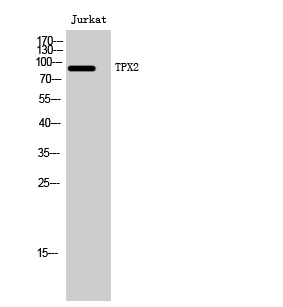
- Western Blot analysis of Jurkat cells using TPX2 Polyclonal Antibody. Secondary antibody(catalog#:RS0002) was diluted at 1:20000 cells nucleus extracted by Minute TM Cytoplasmic and Nuclear Fractionation kit (SC-003,Inventbiotech,MN,USA).

- Immunohistochemistry analysis of paraffin-embedded human brain tissue, using DIL-2 Antibody. The picture on the right is blocked with the synthesized peptide.
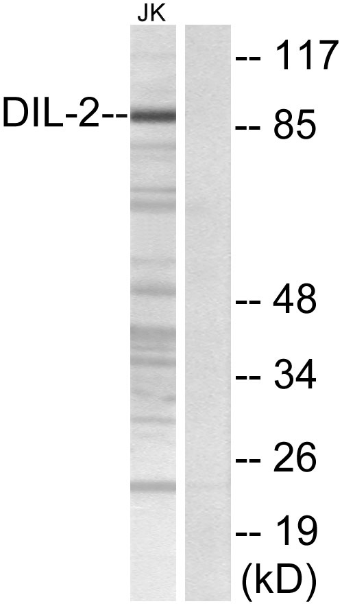
- Western blot analysis of lysates from Jurkat cells, using DIL-2 Antibody. The lane on the right is blocked with the synthesized peptide.
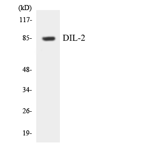
- Western blot analysis of the lysates from HUVECcells using DIL-2 antibody.
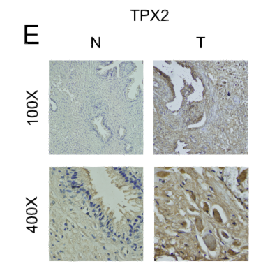
- A Liquid–Liquid Phase Separation-Related Index Associate with Biochemical Recurrence and Tumor Immune Environment of Prostate Cancer Patients INTERNATIONAL JOURNAL OF MOLECULAR SCIENCES Ning Xu IHC Human benign prostatic hyperplasia (BPH) tissue prostate cancer (PCa)cell


