V-ATPase B1 Polyclonal Antibody
- Catalog No.:YT4858
- Applications:WB;IHC;IF;ELISA
- Reactivity:Human;Mouse
- Target:
- V-ATPase B1
- Fields:
- >>Oxidative phosphorylation;>>Metabolic pathways;>>Phagosome;>>mTOR signaling pathway;>>Synaptic vesicle cycle;>>Collecting duct acid secretion;>>Vibrio cholerae infection;>>Epithelial cell signaling in Helicobacter pylori infection;>>Human papillomavirus infection;>>Rheumatoid arthritis
- Gene Name:
- ATP6V1B1
- Protein Name:
- V-type proton ATPase subunit B kidney isoform
- Human Gene Id:
- 525
- Human Swiss Prot No:
- P15313
- Immunogen:
- The antiserum was produced against synthesized peptide derived from human ATP6V1B1. AA range:381-430
- Specificity:
- V-ATPase B1 Polyclonal Antibody detects endogenous levels of V-ATPase B1 protein.
- Formulation:
- Liquid in PBS containing 50% glycerol, 0.5% BSA and 0.02% sodium azide.
- Source:
- Polyclonal, Rabbit,IgG
- Dilution:
- WB 1:500 - 1:2000. IHC 1:100 - 1:300. ELISA: 1:5000.. IF 1:50-200
- Purification:
- The antibody was affinity-purified from rabbit antiserum by affinity-chromatography using epitope-specific immunogen.
- Concentration:
- 1 mg/ml
- Storage Stability:
- -15°C to -25°C/1 year(Do not lower than -25°C)
- Other Name:
- ATP6V1B1;ATP6B1;VATB;VPP3;V-type proton ATPase subunit B; kidney isoform;V-ATPase subunit B 1;Endomembrane proton pump 58 kDa subunit;Vacuolar proton pump subunit B 1
- Observed Band(KD):
- 60kD
- Background:
- This gene encodes a component of vacuolar ATPase (V-ATPase), a multisubunit enzyme that mediates acidification of eukaryotic intracellular organelles. V-ATPase dependent organelle acidification is necessary for such intracellular processes as protein sorting, zymogen activation, receptor-mediated endocytosis, and synaptic vesicle proton gradient generation. V-ATPase is composed of a cytosolic V1 domain and a transmembrane V0 domain. The V1 domain consists of three A and three B subunits, two G subunits plus the C, D, E, F, and H subunits. The V1 domain contains the ATP catalytic site. The V0 domain consists of five different subunits: a, c, c', c'', and d. Additional isoforms of many of the V1 and V0 subunit proteins are encoded by multiple genes or alternatively spliced transcript variants. This encoded protein is one of two V1 domain B subunit isoforms and is found i
- Function:
- disease:Defects in ATP6V1B1 are the cause of distal renal tubular acidosis with deafness (dRTA) [MIM:267300]. Inheritance is autosomal recessive. Patients with recessive dRTA are severely affected, presenting with either acute illness or growth failure at a young age, and bilateral sensorineural deafness. Other features include low serum K(+) due to renal potassium wasting, and elevated urinary calcium. If untreated, this acidosis may result in dissolution of bone, leading to osteomalacia and rickets. Renal deposition of calcium salts (nephrocalcinosis) and renal stone formation commonly occur.,domain:The PDZ-binding motif mediates interactions with SLC9A3R1 and SCL4A7.,function:Non-catalytic subunit of the peripheral V1 complex of vacuolar ATPase. V-ATPase is responsible for acidifying a variety of intracellular compartments in eukaryotic cells.,similarity:Belongs to the ATPase alpha/be
- Subcellular Location:
- Apical cell membrane . Basolateral cell membrane .
- Expression:
- Kidney; localizes to early distal nephron, encompassing thick ascending limbs and distal convoluted tubules (at protein level) (PubMed:29993276, PubMed:16769747). Expressed in the cochlea and endolymphatic sac (PubMed:9916796).
- June 19-2018
- WESTERN IMMUNOBLOTTING PROTOCOL
- June 19-2018
- IMMUNOHISTOCHEMISTRY-PARAFFIN PROTOCOL
- June 19-2018
- IMMUNOFLUORESCENCE PROTOCOL
- September 08-2020
- FLOW-CYTOMEYRT-PROTOCOL
- May 20-2022
- Cell-Based ELISA│解您多样本WB检测之困扰
- July 13-2018
- CELL-BASED-ELISA-PROTOCOL-FOR-ACETYL-PROTEIN
- July 13-2018
- CELL-BASED-ELISA-PROTOCOL-FOR-PHOSPHO-PROTEIN
- July 13-2018
- Antibody-FAQs
- Products Images

- Western Blot analysis of various cells using V-ATPase B1 Polyclonal Antibody. Secondary antibody(catalog#:RS0002) was diluted at 1:20000
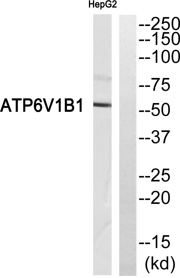
- Western blot analysis of ATP6V1B1 Antibody. The lane on the right is blocked with the ATP6V1B1 peptide.
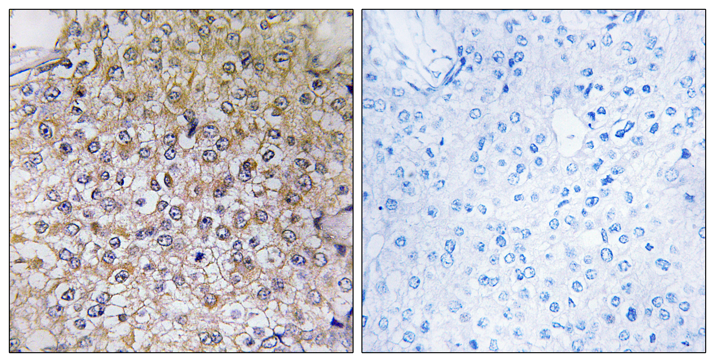
- Immunohistochemistryt analysis of paraffin-embedded human breast carcinoma, using ATP6V1B1 Antibody. The lane on the right is blocked with the ATP6V1B1 peptide.


