VHR Polyclonal Antibody
- Catalog No.:YT5228
- Applications:WB;IHC;IF;ELISA
- Reactivity:Human;Mouse;Rat
- Target:
- VHR
- Fields:
- >>MAPK signaling pathway
- Gene Name:
- DUSP3
- Protein Name:
- Dual specificity protein phosphatase 3
- Human Gene Id:
- 1845
- Human Swiss Prot No:
- P51452
- Mouse Gene Id:
- 72349
- Mouse Swiss Prot No:
- Q9D7X3
- Immunogen:
- The antiserum was produced against synthesized peptide derived from the C-terminal region of human DUSP3. AA range:136-185
- Specificity:
- VHR Polyclonal Antibody detects endogenous levels of VHR protein.
- Formulation:
- Liquid in PBS containing 50% glycerol, 0.5% BSA and 0.02% sodium azide.
- Source:
- Polyclonal, Rabbit,IgG
- Dilution:
- WB 1:500 - 1:2000. IHC: 1:100-300 ELISA: 1:20000.. IF 1:50-200
- Purification:
- The antibody was affinity-purified from rabbit antiserum by affinity-chromatography using epitope-specific immunogen.
- Concentration:
- 1 mg/ml
- Storage Stability:
- -15°C to -25°C/1 year(Do not lower than -25°C)
- Other Name:
- DUSP3;VHR;Dual specificity protein phosphatase 3;Dual specificity protein phosphatase VHR;Vaccinia H1-related phosphatase;VHR
- Observed Band(KD):
- 21kD
- Background:
- The protein encoded by this gene is a member of the dual specificity protein phosphatase subfamily. These phosphatases inactivate their target kinases by dephosphorylating both the phosphoserine/threonine and phosphotyrosine residues. They negatively regulate members of the mitogen-activated protein (MAP) kinase superfamily (MAPK/ERK, SAPK/JNK, p38), which are associated with cellular proliferation and differentiation. Different members of the family of dual specificity phosphatases show distinct substrate specificities for various MAP kinases, different tissue distribution and subcellular localization, and different modes of inducibility of their expression by extracellular stimuli. This gene maps in a region that contains the BRCA1 locus which confers susceptibility to breast and ovarian cancer. Although DUSP3 is expressed in both breast and ovarian tissues, mutation screening in breast ca
- Function:
- catalytic activity:A phosphoprotein + H(2)O = a protein + phosphate.,catalytic activity:Protein tyrosine phosphate + H(2)O = protein tyrosine + phosphate.,function:This protein shows activity both toward tyrosine-protein phosphate as well as with serine-protein phosphate.,similarity:Belongs to the protein-tyrosine phosphatase family. Non-receptor class dual specificity subfamily.,similarity:Contains 1 tyrosine-protein phosphatase domain.,
- Subcellular Location:
- Nucleus .
- Expression:
- Duodenum,Uterus,
- June 19-2018
- WESTERN IMMUNOBLOTTING PROTOCOL
- June 19-2018
- IMMUNOHISTOCHEMISTRY-PARAFFIN PROTOCOL
- June 19-2018
- IMMUNOFLUORESCENCE PROTOCOL
- September 08-2020
- FLOW-CYTOMEYRT-PROTOCOL
- May 20-2022
- Cell-Based ELISA│解您多样本WB检测之困扰
- July 13-2018
- CELL-BASED-ELISA-PROTOCOL-FOR-ACETYL-PROTEIN
- July 13-2018
- CELL-BASED-ELISA-PROTOCOL-FOR-PHOSPHO-PROTEIN
- July 13-2018
- Antibody-FAQs
- Products Images

- Western Blot analysis of HepG2 cells using VHR Polyclonal Antibody. Secondary antibody(catalog#:RS0002) was diluted at 1:20000 cells nucleus extracted by Minute TM Cytoplasmic and Nuclear Fractionation kit (SC-003,Inventbiotech,MN,USA).

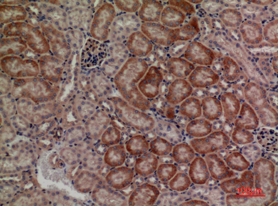
- Immunohistochemical analysis of paraffin-embedded mouse-kidney, antibody was diluted at 1:100

- Western blot analysis of lysate from HepG2 cells, using DUSP3 Antibody.


