CD63 Polyclonal Antibody
- Catalog No.:YT5525
- Applications:IF;WB;IHC;ELISA
- Reactivity:Human;Rat;Mouse;
- Target:
- CD63
- Fields:
- >>Lysosome;>>Proteoglycans in cancer
- Gene Name:
- CD63
- Protein Name:
- CD63 antigen
- Human Gene Id:
- 967
- Human Swiss Prot No:
- P08962
- Mouse Swiss Prot No:
- P41731
- Immunogen:
- The antiserum was produced against synthesized peptide derived from the Internal region of human CD63. AA range:121-170
- Specificity:
- CD63 Polyclonal Antibody detects endogenous levels of CD63 protein.
- Formulation:
- Liquid in PBS containing 50% glycerol, 0.5% BSA and 0.02% sodium azide.
- Source:
- Polyclonal, Rabbit,IgG
- Dilution:
- IF 1:50-200 WB 1:500 - 1:2000. IHC: 1:100-1:300. ELISA: 1:20000. Not yet tested in other applications.
- Purification:
- The antibody was affinity-purified from rabbit antiserum by affinity-chromatography using epitope-specific immunogen.
- Concentration:
- 1 mg/ml
- Storage Stability:
- -15°C to -25°C/1 year(Do not lower than -25°C)
- Other Name:
- CD63;MLA1;TSPAN30;CD63 antigen;Granulophysin;Lysosomal-associated membrane protein 3;LAMP-3;Melanoma-associated antigen ME491;OMA81H;Ocular melanoma-associated antigen;Tetraspanin-30;Tspan-30;CD63
- Observed Band(KD):
- 26,35-65(kD
- Background:
- The protein encoded by this gene is a member of the transmembrane 4 superfamily, also known as the tetraspanin family. Most of these members are cell-surface proteins that are characterized by the presence of four hydrophobic domains. The proteins mediate signal transduction events that play a role in the regulation of cell development, activation, growth and motility. The encoded protein is a cell surface glycoprotein that is known to complex with integrins. It may function as a blood platelet activation marker. Deficiency of this protein is associated with Hermansky-Pudlak syndrome. Also this gene has been associated with tumor progression. Alternative splicing results in multiple transcript variants encoding different protein isoforms. [provided by RefSeq, Apr 2012],
- Function:
- function:This antigen is associated with early stages of melanoma tumor progression. May play a role in growth regulation.,miscellaneous:Lack of expression of CD63 in platelets has been observed in a patient with Hermansky-Pudlak syndrome (HPS). Hermansky-Pudlak syndrome (HPS) is a genetically heterogeneous, rare, autosomal recessive disorder characterized by oculocutaneous albinism, bleeding due to platelet storage pool deficiency, and lysosomal storage defects. This syndrome results from defects of diverse cytoplasmic organelles including melanosomes, platelet dense granules and lysosomes. Ceroid storage in the lungs is associated with pulmonary fibrosis, a common cause of premature death in individuals with HPS.,similarity:Belongs to the tetraspanin (TM4SF) family.,subcellular location:Also found in Weibel-Palade bodies of endothelial cells. Located in platelet dense granules.,tissue
- Subcellular Location:
- Cell membrane ; Multi-pass membrane protein . Lysosome membrane ; Multi-pass membrane protein . Late endosome membrane ; Multi-pass membrane protein . Endosome, multivesicular body . Melanosome . Secreted, extracellular exosome . Cell surface . Also found in Weibel-Palade bodies of endothelial cells (PubMed:10793155). Located in platelet dense granules (PubMed:7682577). Detected in a subset of pre-melanosomes. Detected on intralumenal vesicles (ILVs) within multivesicular bodies (PubMed:21962903). .
- Expression:
- Detected in platelets (at protein level). Dysplastic nevi, radial growth phase primary melanomas, hematopoietic cells, tissue macrophages.
Plasma Exosomal AKR1C3 mRNA Expression Is a Predictive and Prognostic Biomarker in Patients with Metastatic Castration-Resistant Prostate Cancer
Adipose-derived mesenchymal stem cells and extracellular vesicles confer antitumor activity in preclinical treatment of breast cancer. PHARMACOLOGICAL RESEARCH 2020 Apr 30 WB Mouse ADSCs, CD90high ADSC‑EVs,CD90low ADSC‑EVs
Mesenchymal stem cell‑derived extracellular vesicles promote the in vitro proliferation and migration of breast cancer cells through the activation of the ERK pathway. INTERNATIONAL JOURNAL OF ONCOLOGY Int J Oncol. 2019 May;54(5):1843-1852 WB Human 1:1000 hUC-MSC-EVs
Comparative proteomics analysis of microvesicles in human serum for the evaluation of osteoporosis. ELECTROPHORESIS Electrophoresis. 2019 Jul;40(14):1839-1847 WB Human Serum MVs
Periodontal ligament fibroblasts regulate osteoblasts by exosome secretion induced by inflammatory stimuli. ARCHIVES OF ORAL BIOLOGY Arch Oral Biol. 2019 Sep;105:27 WB Human 1:300 hPDLFs
Systematic comparison of exosomal proteomes from human saliva and serum for the detection of lung cancer. ANALYTICA CHIMICA ACTA 2017 Jun 17 WB Human 1:500 saliva exosomes,serum exosomes
Exosomes derived from human umbilical cord mesenchymal stem cells loaded with RVG-Lamp2b and Netrin-1 promotes Schwann cell invasion and migration. TISSUE & CELL Hua Zhang WB Human 1:3000 IMP-H122 cell
Clinical and preclinical evaluation of miR-144-5p as a key target for major depressive disorder. Huanzhong Liu WB Human serum
The value of urinary exosomal lncRNA SNHG16 as a diagnostic biomarker for bladder cancer. MOLECULAR BIOLOGY REPORTS Li Guangyuan WB Human urinary exosomes
Placenta-derived exosomal miR-135a-5p promotes gestational diabetes mellitus pathogenesis by activating PI3K/AKT signalling pathway via SIRT1. JOURNAL OF CELLULAR AND MOLECULAR MEDICINE Jianying Yan WB Human plasma
Exosomes derived from human umbilical cord mesenchymal stem cells loaded with RVG-Lamp2b and Netrin-1 promotes Schwann cell invasion and migration. TISSUE & CELL Hua Zhang WB Human 1:3000 IMP-H122 cell
Extracellular vesicles derived from monomeric α-synuclein-treated microglia ameliorate neuroinflammation by delivery of miRNAs targeting PRAK NEUROSCIENCE LETTERS Na Li WB Mouse 1:1000 BV2 microglia
Bio-nanoparticles loaded with synovial-derived exosomes ameliorate osteoarthritis progression by modifying the oxidative microenvironment JOURNAL OF NANOBIOTECHNOLOGY Cao Haifei WB Mouse synovial fibroblasts
Engineered nanovesicles from activated neutrophils with enriched bactericidal proteins have molecular debridement ability and promote infectious wound healing Burns & Trauma Jin Hangfei WB Human Neutrophils
Attenuation of Cardiomyocyte Hypertrophy Via Inhalation of Isosteviol-Loaded Exosome in Mice PHARMACEUTICAL CHEMISTRY JOURNAL Guo Haihua WB Human HEK293 cell
- June 19-2018
- WESTERN IMMUNOBLOTTING PROTOCOL
- June 19-2018
- IMMUNOHISTOCHEMISTRY-PARAFFIN PROTOCOL
- June 19-2018
- IMMUNOFLUORESCENCE PROTOCOL
- September 08-2020
- FLOW-CYTOMEYRT-PROTOCOL
- May 20-2022
- Cell-Based ELISA│解您多样本WB检测之困扰
- July 13-2018
- CELL-BASED-ELISA-PROTOCOL-FOR-ACETYL-PROTEIN
- July 13-2018
- CELL-BASED-ELISA-PROTOCOL-FOR-PHOSPHO-PROTEIN
- July 13-2018
- Antibody-FAQs
- Products Images
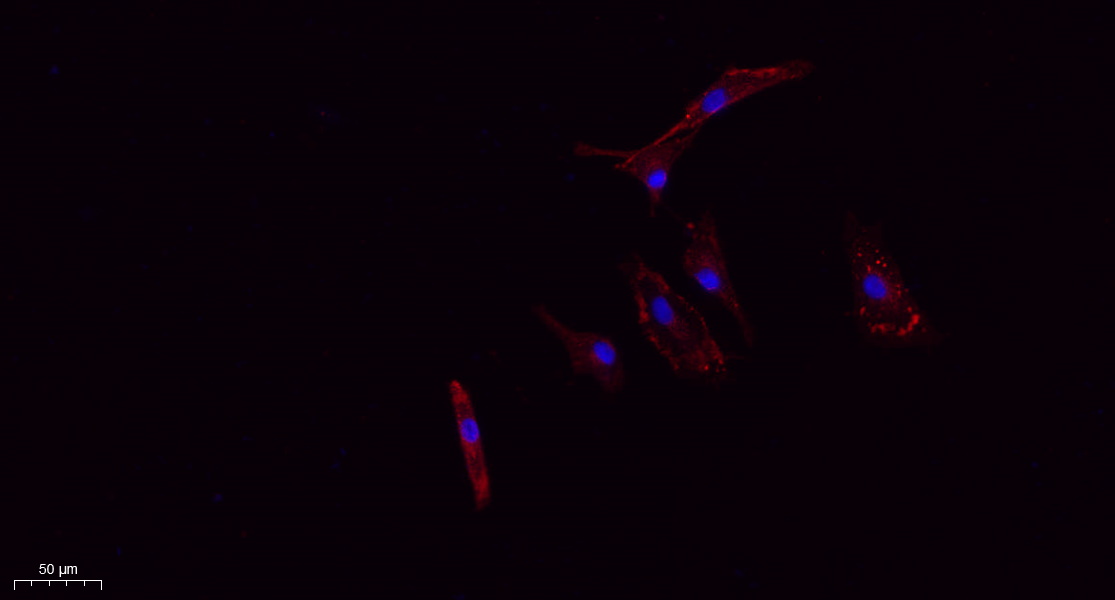
- Immunofluorescence analysis of A549. 1,primary Antibody(red) was diluted at 1:200(4°C overnight). 2, Goat Anti Rabbit IgG (H&L) - Alexa Fluor 594 Secondary antibody was diluted at 1:1000(room temperature, 50min).3, Picture B: DAPI(blue) 10min.
.jpg)
- Zhang, Z., Xu, R., Yang, Y. et al. Micro/nano-textured hierarchical titanium topography promotes exosome biogenesis and secretion to improve osseointegration. J Nanobiotechnol 19, 78 (2021).
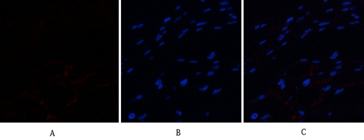
- Immunofluorescence analysis of human-stomach-cancer tissue. 1,CD63 Polyclonal Antibody(red) was diluted at 1:200(4°C,overnight). 2, Cy3 labled Secondary antibody was diluted at 1:300(room temperature, 50min).3, Picture B: DAPI(blue) 10min. Picture A:Target. Picture B: DAPI. Picture C: merge of A+B
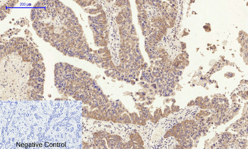
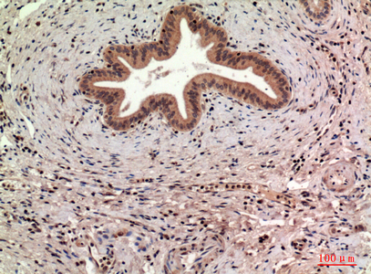
- Immunohistochemical analysis of paraffin-embedded Human-liver-cancer tissue. 1,CD63 Polyclonal Antibody was diluted at 1:200(4°C,overnight). 2, Sodium citrate pH 6.0 was used for antibody retrieval(>98°C,20min). 3,Secondary antibody was diluted at 1:200(room tempeRature, 30min). Negative control was used by secondary antibody only.
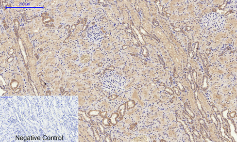
- Immunohistochemical analysis of paraffin-embedded Human-kidney tissue. 1,CD63 Polyclonal Antibody was diluted at 1:200(4°C,overnight). 2, Sodium citrate pH 6.0 was used for antibody retrieval(>98°C,20min). 3,Secondary antibody was diluted at 1:200(room tempeRature, 30min). Negative control was used by secondary antibody only.
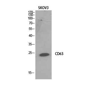
- Western Blot analysis of SKOV3 cells using CD63 Polyclonal Antibody. Secondary antibody(catalog#:RS0002) was diluted at 1:20000
.jpg)
- Immunohistochemical analysis of paraffin-embedded human-liver, antibody was diluted at 1:100
- Immunohistochemical analysis of paraffin-embedded human-liver, antibody was diluted at 1:100

- Immunohistochemical analysis of paraffin-embedded human-lung, antibody was diluted at 1:100


