PI 3-Kinase p110δ Polyclonal Antibody
- Catalog No.:YT5537
- Applications:WB;IHC;IF;ELISA
- Reactivity:Human;Mouse
- Target:
- PI 3-Kinase p110δ
- Fields:
- >>Inositol phosphate metabolism;>>Metabolic pathways;>>EGFR tyrosine kinase inhibitor resistance;>>Endocrine resistance;>>Platinum drug resistance;>>ErbB signaling pathway;>>Ras signaling pathway;>>Rap1 signaling pathway;>>cAMP signaling pathway;>>Chemokine signaling pathway;>>HIF-1 signaling pathway;>>FoxO signaling pathway;>>Phosphatidylinositol signaling system;>>Sphingolipid signaling pathway;>>Phospholipase D signaling pathway;>>Autophagy - animal;>>mTOR signaling pathway;>>PI3K-Akt signaling pathway;>>AMPK signaling pathway;>>Apoptosis;>>Longevity regulating pathway;>>Longevity regulating pathway - multiple species;>>Cellular senescence;>>Axon guidance;>>VEGF signaling pathway;>>Osteoclast differentiation;>>Focal adhesion;>>Signaling pathways regulating pluripotency of stem cells;>>Platelet activation;>>Neutrophil extracellular trap formation;>>Toll-like receptor signaling pathway;>>C-type lectin receptor signaling pathway;>>JAK-STAT signaling pathway;>>Natural killer cell mediat
- Gene Name:
- PIK3CD
- Protein Name:
- Phosphatidylinositol 4,5-bisphosphate 3-kinase catalytic subunit delta isoform
- Human Gene Id:
- 5293
- Human Swiss Prot No:
- O00329
- Mouse Gene Id:
- 18707
- Mouse Swiss Prot No:
- O35904
- Immunogen:
- The antiserum was produced against synthesized peptide derived from the N-terminal region of human PIK3CD. AA range:41-90
- Specificity:
- PI 3-Kinase p110δ Polyclonal Antibody detects endogenous levels of PI 3-Kinase p110δ protein.
- Formulation:
- Liquid in PBS containing 50% glycerol, 0.5% BSA and 0.02% sodium azide.
- Source:
- Polyclonal, Rabbit,IgG
- Dilution:
- WB 1:500 - 1:2000. IHC: 1:100-1:300. ELISA: 1:20000.. IF 1:50-200
- Purification:
- The antibody was affinity-purified from rabbit antiserum by affinity-chromatography using epitope-specific immunogen.
- Concentration:
- 1 mg/ml
- Storage Stability:
- -15°C to -25°C/1 year(Do not lower than -25°C)
- Other Name:
- PIK3CD;Phosphatidylinositol 4,5-bisphosphate 3-kinase catalytic subunit delta isoform;PI3-kinase subunit delta;PI3K-delta;PI3Kdelta;PtdIns-3-kinase subunit delta;Phosphatidylinositol 4,5-bisphosphate 3-kinase 110 kDa catalytic subunit delta;PtdIns-3-kinase subunit p110-delta;p110delta
- Observed Band(KD):
- 120kD
- Background:
- Phosphoinositide 3-kinases (PI3Ks) phosphorylate inositol lipids and are involved in the immune response. The protein encoded by this gene is a class I PI3K found primarily in leukocytes. Like other class I PI3Ks (p110-alpha p110-beta, and p110-gamma), the encoded protein binds p85 adapter proteins and GTP-bound RAS. However, unlike the other class I PI3Ks, this protein phosphorylates itself, not p85 protein.[provided by RefSeq, Jul 2010],
- Function:
- catalytic activity:ATP + 1-phosphatidyl-1D-myo-inositol 4,5-bisphosphate = ADP + 1-phosphatidyl-1D-myo-inositol 3,4,5-trisphosphate.,pathway:Phospholipid metabolism; phosphatidylinositol phosphate biosynthesis.,PTM:Autophosphorylation on Ser-1039 results in the almost complete inactivation of the lipid kinase activity.,similarity:Belongs to the PI3/PI4-kinase family.,similarity:Contains 1 PI3K/PI4K domain.,subunit:Heterodimer of a p110 (catalytic) and a p85 (regulatory) subunit. Interacts with ERAS.,tissue specificity:Expressed predominantly in leukocytes.,
- Subcellular Location:
- Cytoplasm .
- Expression:
- In humans, the highest levels of expression are seen in peripheral blood mononuclear cells, spleen, and thymus, and low levels of expression in testes, uterus, colon, and small intestine but not in other tissues examined including prostate, heart, brain, and liver (PubMed:9235916). Isoform 2 is expressed in normal thymus, lung and spleen tissues, and is detected at low levels in normal lysates from colon and ovarian biopsies, at elevated levels in lysates from colorectal tumors and is abundantly expressed in some ovarian tumors (at protein level). Both isoform 1 and isoform 2 are widely expressed. Isoform 1 is expressed predominantly in leukocytes.
- June 19-2018
- WESTERN IMMUNOBLOTTING PROTOCOL
- June 19-2018
- IMMUNOHISTOCHEMISTRY-PARAFFIN PROTOCOL
- June 19-2018
- IMMUNOFLUORESCENCE PROTOCOL
- September 08-2020
- FLOW-CYTOMEYRT-PROTOCOL
- May 20-2022
- Cell-Based ELISA│解您多样本WB检测之困扰
- July 13-2018
- CELL-BASED-ELISA-PROTOCOL-FOR-ACETYL-PROTEIN
- July 13-2018
- CELL-BASED-ELISA-PROTOCOL-FOR-PHOSPHO-PROTEIN
- July 13-2018
- Antibody-FAQs
- Products Images
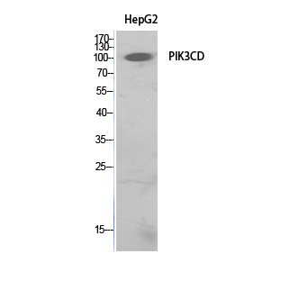
- Western Blot analysis of HepG2 cells using PI 3-Kinase p110δ Polyclonal Antibody. Secondary antibody(catalog#:RS0002) was diluted at 1:20000
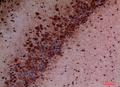
- Immunohistochemical analysis of paraffin-embedded mouse-brain, antibody was diluted at 1:100


