M-CSF Polyclonal Antibody
- Catalog No.:YT5553
- Applications:WB;IHC;IF;ELISA
- Reactivity:Human;Rat;Mouse;
- Target:
- M-CSF
- Fields:
- >>MAPK signaling pathway;>>Ras signaling pathway;>>Rap1 signaling pathway;>>Cytokine-cytokine receptor interaction;>>Viral protein interaction with cytokine and cytokine receptor;>>PI3K-Akt signaling pathway;>>Osteoclast differentiation;>>Hematopoietic cell lineage;>>TNF signaling pathway;>>Alzheimer disease;>>Pathways of neurodegeneration - multiple diseases;>>Rheumatoid arthritis
- Gene Name:
- CSF1
- Protein Name:
- Macrophage colony-stimulating factor 1
- Human Gene Id:
- 1435
- Human Swiss Prot No:
- P09603
- Mouse Swiss Prot No:
- P07141
- Immunogen:
- The antiserum was produced against synthesized peptide derived from the C-terminal region of human CSF1. AA range:505-554
- Specificity:
- M-CSF Polyclonal Antibody detects endogenous levels of M-CSF protein.
- Formulation:
- Liquid in PBS containing 50% glycerol, 0.5% BSA and 0.02% sodium azide.
- Source:
- Polyclonal, Rabbit,IgG
- Dilution:
- WB 1:500 - 1:2000. IHC: 1:100-1:300. ELISA: 1:10000.. IF 1:50-200
- Purification:
- The antibody was affinity-purified from rabbit antiserum by affinity-chromatography using epitope-specific immunogen.
- Concentration:
- 1 mg/ml
- Storage Stability:
- -15°C to -25°C/1 year(Do not lower than -25°C)
- Other Name:
- CSF1;Macrophage colony-stimulating factor 1;CSF-1;M-CSF;MCSF;Lanimostim
- Observed Band(KD):
- 48kD
- Background:
- The protein encoded by this gene is a cytokine that controls the production, differentiation, and function of macrophages. The active form of the protein is found extracellularly as a disulfide-linked homodimer, and is thought to be produced by proteolytic cleavage of membrane-bound precursors. The encoded protein may be involved in development of the placenta. Alternate splicing results in multiple transcript variants. [provided by RefSeq, Sep 2011],
- Function:
- function:Granulocyte/macrophage colony-stimulating factors are cytokines that act in hematopoiesis by controlling the production, differentiation, and function of 2 related white cell populations of the blood, the granulocytes and the monocytes-macrophages. CSF-1 induces cells of the monocyte/macrophage lineage. It plays a role in immunological defenses, bone metabolism, lipoproteins clearance, fertility and pregnancy.,PTM:Glycosylation and proteolytic cleavage yield different soluble forms. A high molecular weight soluble form is a proteoglycan containing chondroitin sulfate.,PTM:Isoform 1 is N- and O-glycosylated. Isoform 3 is N-glycosylated.,subunit:Homodimer or heterodimer; disulfide-linked.,
- Subcellular Location:
- Cell membrane ; Single-pass type I membrane protein .; [Processed macrophage colony-stimulating factor 1]: Secreted, extracellular space.
- Expression:
- Endometrium,Kidney,Pancreatic carcinoma,T lymphoblast,Trophoblast,Urine,
- June 19-2018
- WESTERN IMMUNOBLOTTING PROTOCOL
- June 19-2018
- IMMUNOHISTOCHEMISTRY-PARAFFIN PROTOCOL
- June 19-2018
- IMMUNOFLUORESCENCE PROTOCOL
- September 08-2020
- FLOW-CYTOMEYRT-PROTOCOL
- May 20-2022
- Cell-Based ELISA│解您多样本WB检测之困扰
- July 13-2018
- CELL-BASED-ELISA-PROTOCOL-FOR-ACETYL-PROTEIN
- July 13-2018
- CELL-BASED-ELISA-PROTOCOL-FOR-PHOSPHO-PROTEIN
- July 13-2018
- Antibody-FAQs
- Products Images

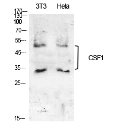
- Western Blot analysis of NIH-3T3, Hela cells using M-CSF Polyclonal Antibody. Secondary antibody(catalog#:RS0002) was diluted at 1:20000
.jpg)
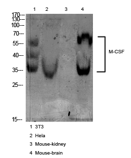
- Western Blot analysis of 3T3 cells using M-CSF Polyclonal Antibody. Secondary antibody(catalog#:RS0002) was diluted at 1:20000
.jpg)
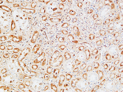
- Immunohistochemical analysis of paraffin-embedded human-kidney, antibody was diluted at 1:100

- Western blot analysis of various cell Lysate, antibody was diluted at 1:1000. Secondary antibody(catalog#:RS0002) was diluted at 1:20000

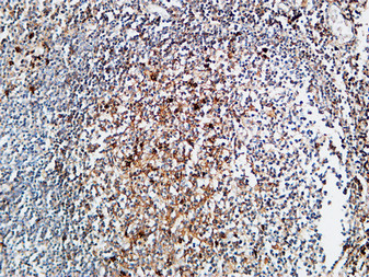
- Immunohistochemical analysis of paraffin-embedded Human Amygdala. 1, Antibody was diluted at 1:200(4° overnight). 2, High-pressure and temperature EDTA, pH8.0 was used for antigen retrieval. 3,Secondary antibody was diluted at 1:200(room temperature, 30min).

- Immunohistochemical analysis of paraffin-embedded Human kidney. 1, Antibody was diluted at 1:200(4° overnight). 2, High-pressure and temperature EDTA, pH8.0 was used for antigen retrieval. 3,Secondary antibody was diluted at 1:200(room temperature, 30min).


