SorLA Monoclonal Antibody
- Catalog No.:YM0591
- Applications:WB;IHC;IF;ELISA
- Reactivity:Human
- Target:
- SorLA
- Gene Name:
- SORL1
- Protein Name:
- Sortilin-related receptor
- Human Gene Id:
- 6653
- Human Swiss Prot No:
- Q92673
- Mouse Swiss Prot No:
- O88307
- Immunogen:
- Purified recombinant fragment of human SorLA expressed in E. Coli.
- Specificity:
- SorLA Monoclonal Antibody detects endogenous levels of SorLA protein.
- Formulation:
- Liquid in PBS containing 50% glycerol, 0.5% BSA and 0.02% sodium azide.
- Source:
- Monoclonal, Mouse
- Dilution:
- WB 1:500 - 1:2000. IHC 1:200 - 1:1000. IF 1:200 - 1:1000. ELISA: 1:10000. Not yet tested in other applications.
- Purification:
- Affinity purification
- Storage Stability:
- -15°C to -25°C/1 year(Do not lower than -25°C)
- Other Name:
- SORL1;C11orf32;Sortilin-related receptor;Low-density lipoprotein receptor relative with 11 ligand-binding repeats;LDLR relative with 11 ligand-binding repeats;LR11;SorLA-1;Sorting protein-related receptor containing LDLR class A repe
- Molecular Weight(Da):
- 248kD
- References:
- 1. Scherzer CR. Offe K. Gearing M. et al. Arch Neurol. 2004, Aug, 61(8):1200-5.
2. Gabrielsson BG. Olofsson LE. Sjogren A. et al. Obes Res. 2005, Apr, 13(4):649-52.
3. Shah S. Yu G. Mol Interv
- Background:
- This gene encodes a mosaic protein that belongs to at least two families: the vacuolar protein sorting 10 (VPS10) domain-containing receptor family, and the low density lipoprotein receptor (LDLR) family. The encoded protein also contains fibronectin type III repeats and an epidermal growth factor repeat. The encoded preproprotein is proteolytically processed to generate the mature receptor, which likely plays roles in endocytosis and sorting. Mutations in this gene may be associated with Alzheimer's disease. [provided by RefSeq, Feb 2016],
- Function:
- disease:Genetic variations in SORL1 may be associated with increased risk for late onset Alzheimer disease (AD). Alzheimer disease is a neurodegenerative disorder characterized by progressive dementia, loss of cognitive abilities, and deposition of fibrillar amyloid proteins as intraneuronal neurofibrillary tangles, extracellular amyloid plaques and vascular amyloid deposits. The major constituent of these plaques is the neurotoxic amyloid-beta-APP 40-42 peptide (s), derived proteolytically from the transmembrane precursor protein APP by sequential secretase processing. The cytotoxic C-terminal fragments (CTFs) and the caspase-cleaved products such as C31 derived from APP, are also implicated in neuronal death.,function:Likely to be a multifunctional endocytic receptor, that may be implicated in the uptake of lipoproteins and of proteases. Binds LDL, the major cholesterol-carrying lipopr
- Subcellular Location:
- Golgi apparatus membrane ; Single-pass type I membrane protein . Golgi apparatus, trans-Golgi network membrane ; Single-pass type I membrane protein . Endosome membrane ; Single-pass type I membrane protein . Early endosome membrane ; Single-pass type I membrane protein . Recycling endosome membrane ; Single-pass type I membrane protein . Endoplasmic reticulum membrane ; Single-pass type I membrane protein . Endosome, multivesicular body membrane ; Single-pass type I membrane protein . Cell membrane ; Single-pass type I membrane protein . Cytoplasmic vesicle, secretory vesicle membrane ; Single-pass type I membrane protein . Secreted . Mostly intracellular, predominantly in the trans-Golgi network (TGN) and in endosome, as well as in endosome-to-TGN retrograde vesicles; found at low levels
- Expression:
- Highly expressed in brain (at protein level) (PubMed:9157966, PubMed:16174740, PubMed:21147781). Most abundant in the cerebellum, cerebral cortex and occipital pole; low levels in the putamen and thalamus (PubMed:9157966, PubMed:16174740). Expression is significantly reduced in the frontal cortex of patients suffering from Alzheimer disease (PubMed:16174740). Also expressed in spinal cord, spleen, testis, prostate, ovary, thyroid and lymph nodes (PubMed:9157966, PubMed:8940146).
- June 19-2018
- WESTERN IMMUNOBLOTTING PROTOCOL
- June 19-2018
- IMMUNOHISTOCHEMISTRY-PARAFFIN PROTOCOL
- June 19-2018
- IMMUNOFLUORESCENCE PROTOCOL
- September 08-2020
- FLOW-CYTOMEYRT-PROTOCOL
- May 20-2022
- Cell-Based ELISA│解您多样本WB检测之困扰
- July 13-2018
- CELL-BASED-ELISA-PROTOCOL-FOR-ACETYL-PROTEIN
- July 13-2018
- CELL-BASED-ELISA-PROTOCOL-FOR-PHOSPHO-PROTEIN
- July 13-2018
- Antibody-FAQs
- Products Images

- Western Blot analysis using SorLA Monoclonal Antibody against truncated SorLA recombinant protein.

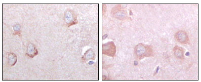
- Immunohistochemistry analysis of paraffin-embedded human cerebrum tissues with DAB staining using SorLA Monoclonal Antibody.

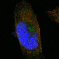
- Confocal immunofluorescence analysis of PANC-1 cells using SorLA Monoclonal Antibody (green). Red: Actin filaments have been labeled with Alexa Fluor-555 phalloidin. Blue: DRAQ5 fluorescent DNA dye.


