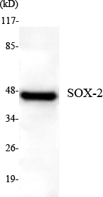
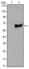
- Target:
- SOX-2
- Fields:
- >>Hippo signaling pathway;>>Signaling pathways regulating pluripotency of stem cells
- Gene Name:
- sox2
- Human Gene Id:
- 20674
- Human Swiss Prot No:
- P48431
- Mouse Swiss Prot No:
- P48432
- Immunogen:
- Purified recombinant mouse Sox2 protein fragments expressed in E.coli
- Specificity:
- This antibody detects endogenous levels of Sox2 and does not cross-react with related proteins.
- Formulation:
- Liquid in PBS containing 50% glycerol, 0.5% BSA and 0.02% sodium azide.
- Source:
- Monoclonal, Mouse
- Dilution:
- wb 1:1000 icc 1:150
- Purification:
- The antibody was affinity-purified from mouse ascites by affinity-chromatography using epitope-specific immunogen.
- Concentration:
- 1 mg/ml
- Storage Stability:
- -15°C to -25°C/1 year(Do not lower than -25°C)
- Other Name:
- ANOP3;cb236;Delta EF2a;lcc;MCOPS3;MGC148683;MGC2413;RGD1565646;Sex determining region Y box 2;SOX 2;Sox2;SOX2_HUMAN;SRY (sex determining region Y) box 2;SRY box containing gene 2;SRY related HMG box 2;SRY related HMG box gene 2;SRY-box 2;Transcription factor SOX 2;Transcription factor SOX-2;ysb.
- Observed Band(KD):
- 35kD
- Background:
- SRY-box 2(SOX2) Homo sapiens This intronless gene encodes a member of the SRY-related HMG-box (SOX) family of transcription factors involved in the regulation of embryonic development and in the determination of cell fate. The product of this gene is required for stem-cell maintenance in the central nervous system, and also regulates gene expression in the stomach. Mutations in this gene have been associated with optic nerve hypoplasia and with syndromic microphthalmia, a severe form of structural eye malformation. This gene lies within an intron of another gene called SOX2 overlapping transcript (SOX2OT). [provided by RefSeq, Jul 2008],
- Function:
- disease:Defects in SOX2 are the cause of microphthalmia syndromic type 3 (MCOPS3) [MIM:206900]. Microphthalmia is a clinically heterogeneous disorder of eye formation, ranging from small size of a single eye to complete bilateral absence of ocular tissues (anophthalmia). In many cases, microphthalmia/anophthalmia occurs in association with syndromes that include non-ocular abnormalities. MCOPS3 is characterized by the rare association of malformations including uni- or bilateral anophthalmia or microphthalmia, and esophageal atresia with trachoesophageal fistula.,function:Transcription factor that forms a trimeric complex with OCT4 on DNA and controls the expression of a number of genes involved in embryonic development such as YES1, FGF4, UTF1 and ZFP206. Critical for early embryogenesis and for embryonic stem cell pluripotency.,online information:Sox2 entry,PTM:Sumoylation inhibits bin
- Subcellular Location:
- Nucleus speckle . Cytoplasm . Nucleus . Acetylation contributes to its nuclear localization and deacetylation by HDAC3 induces a cytoplasmic delocalization (By similarity). Colocalizes in the nucleus with ZNF208 isoform KRAB-O and tyrosine hydroxylase (TH) (By similarity). Colocalizes with SOX6 in speckles. Colocalizes with CAML in the nucleus (By similarity). Nuclear import is facilitated by XPO4, a protein that usually acts as a nuclear export signal receptor (By similarity). .
- Expression:
- Fetal brain,Lung,Retina,
Single cell derived spheres of umbilical cord mesenchymal stem cells enhance cell stemness properties, survival ability and therapeutic potential on liver failure. BIOMATERIALS 2019 Oct 21 WB Human Umbilical cord mesenchymal stem cells (UCMSCs)
- June 19-2018
- WESTERN IMMUNOBLOTTING PROTOCOL
- June 19-2018
- IMMUNOHISTOCHEMISTRY-PARAFFIN PROTOCOL
- June 19-2018
- IMMUNOFLUORESCENCE PROTOCOL
- September 08-2020
- FLOW-CYTOMEYRT-PROTOCOL
- May 20-2022
- Cell-Based ELISA│解您多样本WB检测之困扰
- July 13-2018
- CELL-BASED-ELISA-PROTOCOL-FOR-ACETYL-PROTEIN
- July 13-2018
- CELL-BASED-ELISA-PROTOCOL-FOR-PHOSPHO-PROTEIN
- July 13-2018
- Antibody-FAQs
- Products Images
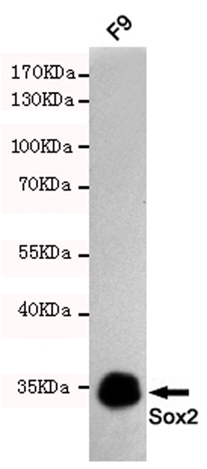
- Western blot detection of Sox2 in F9 cell lysates using Sox2 mouse mAb (1:1000 diluted).Predicted band size:35KDa.Observed band size:35KDa.
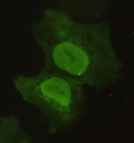
- Immunocytochemistry of COS7 cells using anti-Sox2 mouse mAb diluted 1:150.
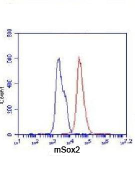
- Flow Cytometry analysis of F9 cells stained with Sox2 (red, 1/100 dilution), followed by FITC-conjugated goat anti-mouse IgG. Blue line histogram represents the isotype control, normal mouse IgG.
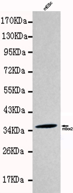
- Western blot detection of Sox2 in mES cell lysates using Sox2 antibody(1:1000 diluted).Predicted band size:35KDa,Observed band size:35KDa.Kindly provided by Dr. Qintong Li at the College of Life Sciences, Sichuan University
