SLP-76 (phospho Tyr128) Polyclonal Antibody
- Catalog No.:YP0801
- Applications:WB;IHC;IF;ELISA
- Reactivity:Human;Mouse;Rat
- Target:
- SLP-76
- Fields:
- >>Rap1 signaling pathway;>>Osteoclast differentiation;>>Platelet activation;>>Natural killer cell mediated cytotoxicity;>>T cell receptor signaling pathway;>>Fc epsilon RI signaling pathway;>>Yersinia infection
- Gene Name:
- LCP2
- Protein Name:
- Lymphocyte cytosolic protein 2
- Human Gene Id:
- 3937
- Human Swiss Prot No:
- Q13094
- Mouse Gene Id:
- 16822
- Mouse Swiss Prot No:
- Q60787
- Immunogen:
- The antiserum was produced against synthesized peptide derived from human SLP-76 around the phosphorylation site of Tyr128. AA range:94-143
- Specificity:
- Phospho-SLP-76 (Y128) Polyclonal Antibody detects endogenous levels of SLP-76 protein only when phosphorylated at Y128.
- Formulation:
- Liquid in PBS containing 50% glycerol, 0.5% BSA and 0.02% sodium azide.
- Source:
- Polyclonal, Rabbit,IgG
- Dilution:
- WB 1:500 - 1:2000. IHC 1:100 - 1:300. ELISA: 1:5000.. IF 1:50-200
- Purification:
- The antibody was affinity-purified from rabbit antiserum by affinity-chromatography using epitope-specific immunogen.
- Concentration:
- 1 mg/ml
- Storage Stability:
- -15°C to -25°C/1 year(Do not lower than -25°C)
- Other Name:
- LCP2;Lymphocyte cytosolic protein 2;SH2 domain-containing leukocyte protein of 76 kDa;SLP-76 tyrosine phosphoprotein;SLP76
- Observed Band(KD):
- 75kD
- Background:
- SLP-76 was originally identified as a substrate of the ZAP-70 protein tyrosine kinase following T cell receptor (TCR) ligation in the leukemic T cell line Jurkat. The SLP-76 locus has been localized to human chromosome 5q33 and the gene structure has been partially characterized in mice. The human and murine cDNAs both encode 533 amino acid proteins that are 72% identical and comprised of three modular domains. The NH2-terminus contains an acidic region that includes a PEST domain and several tyrosine residues which are phosphorylated following TCR ligation. SLP-76 also contains a central proline-rich domain and a COOH-terminal SH2 domain. A number of additional proteins have been identified that associate with SLP-76 both constitutively and inducibly following receptor ligation, supporting the notion that SLP-76 functions as an adaptor or scaffold protein. Studies using SLP-76 deficient T c
- Function:
- domain:The SH2 domain mediates interaction with SHB.,function:Involved in T-cell antigen receptor mediated signaling.,PTM:Phosphorylated after T-cell receptor activation by ZAP-70.,similarity:Contains 1 SAM (sterile alpha motif) domain.,similarity:Contains 1 SH2 domain.,subunit:Interacts with SLA. Interacts with CBLB (By similarity). Interacts with the adapter proteins GRB2 and FYB. Interacts with SHB. Interacts with PRAM1.,tissue specificity:Highly expressed in spleen, thymus, and peripheral blood leukocytes. Highly expressed also in T-cell and monocytic cell lines, expressed at lower level in B-cell lines. Not detected in fibroblast or neuroblasatoma cell lines.,
- Subcellular Location:
- Cytoplasm .
- Expression:
- Highly expressed in spleen, thymus and peripheral blood leukocytes. Highly expressed also in T-cell and monocytic cell lines, expressed at lower level in B-cell lines. Not detected in fibroblast or neuroblastoma cell lines.
- June 19-2018
- WESTERN IMMUNOBLOTTING PROTOCOL
- June 19-2018
- IMMUNOHISTOCHEMISTRY-PARAFFIN PROTOCOL
- June 19-2018
- IMMUNOFLUORESCENCE PROTOCOL
- September 08-2020
- FLOW-CYTOMEYRT-PROTOCOL
- May 20-2022
- Cell-Based ELISA│解您多样本WB检测之困扰
- July 13-2018
- CELL-BASED-ELISA-PROTOCOL-FOR-ACETYL-PROTEIN
- July 13-2018
- CELL-BASED-ELISA-PROTOCOL-FOR-PHOSPHO-PROTEIN
- July 13-2018
- Antibody-FAQs
- Products Images

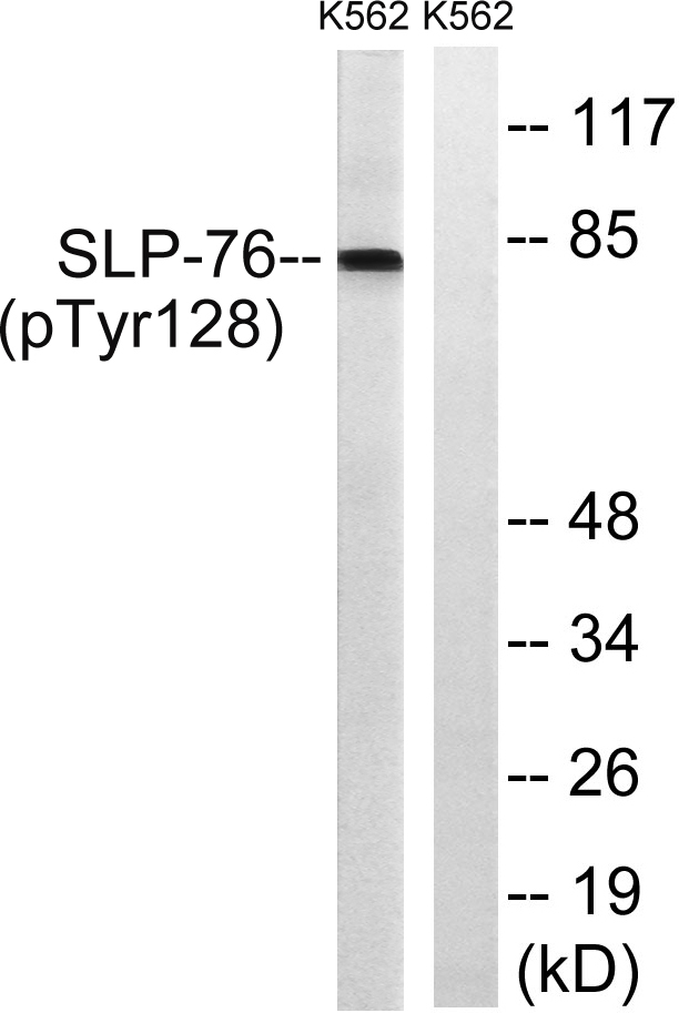
- Western Blot analysis of K562 cells using Phospho-SLP-76 (Y128) Polyclonal Antibody

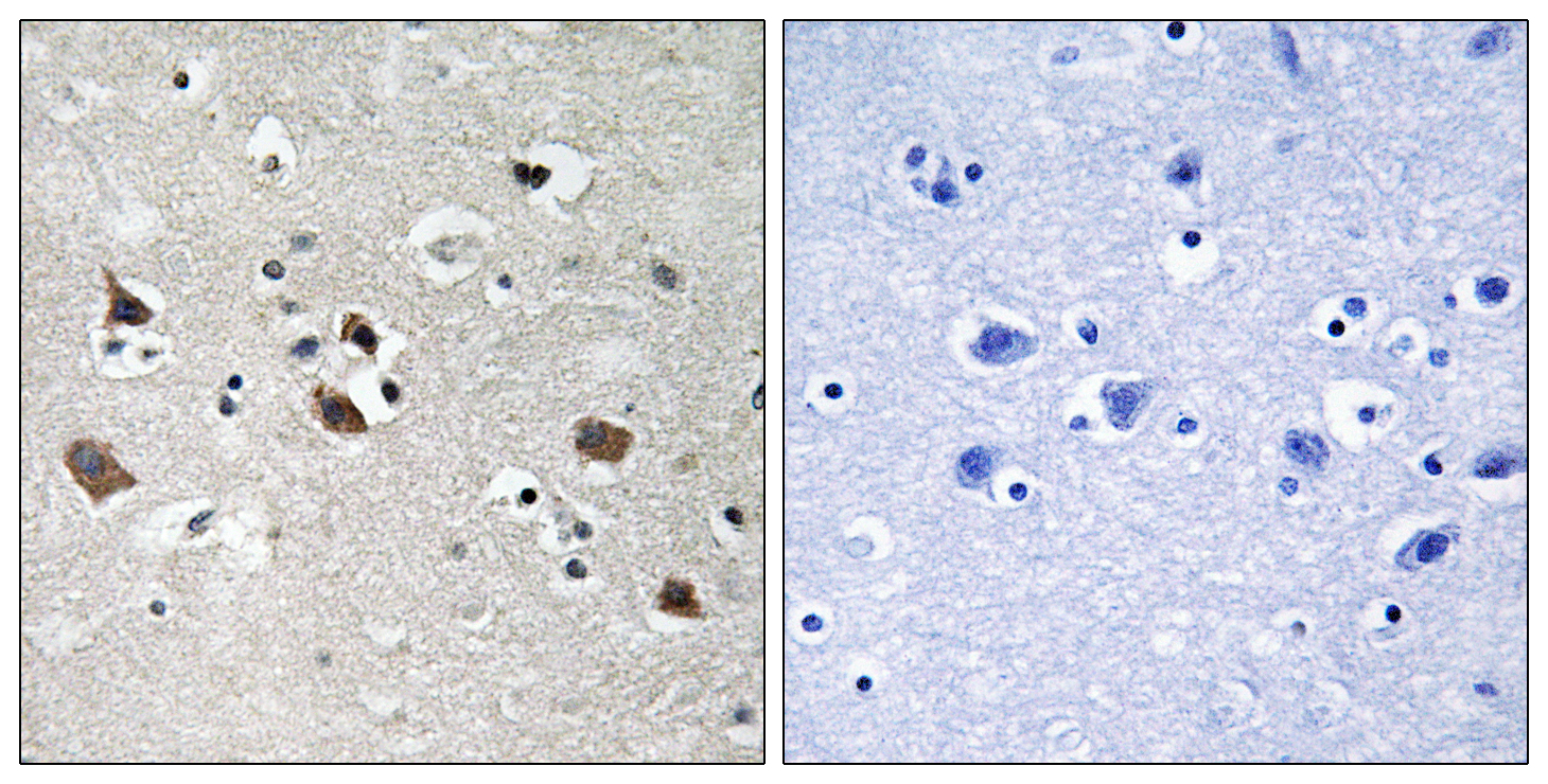
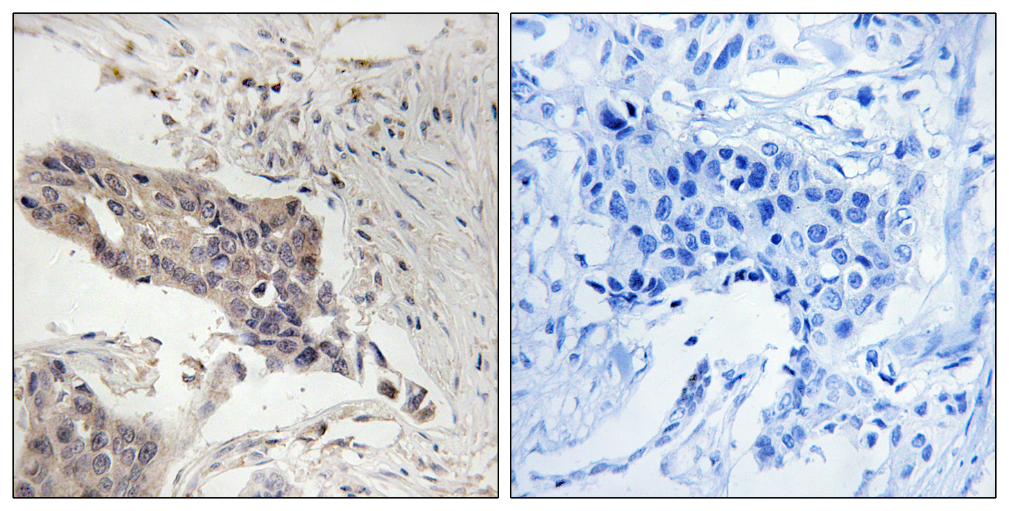
- Immunohistochemistry analysis of paraffin-embedded human brain, using SLP-76 (Phospho-Tyr128) Antibody. The picture on the right is blocked with the phospho peptide.

- Western blot analysis of lysates from K562 cells treated with Na3VO4 0.3nM 40', using SLP-76 (Phospho-Tyr128) Antibody. The lane on the right is blocked with the phospho peptide.


