IRAK-2 Polyclonal Antibody
- Catalog No.:YT2392
- Applications:WB;IHC
- Reactivity:Human;Rat;Mouse;
- Target:
- IRAK-2
- Fields:
- >>Neurotrophin signaling pathway;>>Tuberculosis
- Gene Name:
- IRAK2
- Protein Name:
- Interleukin-1 receptor-associated kinase-like 2
- Human Gene Id:
- 3656
- Human Swiss Prot No:
- O43187
- Mouse Swiss Prot No:
- Q8CFA1
- Immunogen:
- Synthesized peptide derived from the Internal region of human IRAK-2.
- Specificity:
- IRAK-2 Polyclonal Antibody detects endogenous levels of IRAK-2 protein.
- Formulation:
- Liquid in PBS containing 50% glycerol, 0.5% BSA and 0.02% sodium azide.
- Source:
- Polyclonal, Rabbit,IgG
- Dilution:
- WB 1:500-2000;IHC 1:50-300
- Purification:
- The antibody was affinity-purified from rabbit antiserum by affinity-chromatography using epitope-specific immunogen.
- Concentration:
- 1 mg/ml
- Storage Stability:
- -15°C to -25°C/1 year(Do not lower than -25°C)
- Other Name:
- IRAK2;Interleukin-1 receptor-associated kinase-like 2;IRAK-2
- Observed Band(KD):
- 70kD
- Background:
- IRAK2 encodes the interleukin-1 receptor-associated kinase 2, one of two putative serine/threonine kinases that become associated with the interleukin-1 receptor (IL1R) upon stimulation. IRAK2 is reported to participate in the IL1-induced upregulation of NF-kappaB. [provided by RefSeq, Jul 2008],
- Function:
- caution:Asn-335 is present instead of the conserved Asp which is expected to be an active site residue. This enzyme has been shown to be catalytically inactive.,domain:The protein kinase domain is predicted to be catalytically inactive.,function:Binds to the IL-1 type I receptor following IL-1 engagement, triggering intracellular signaling cascades leading to transcriptional up-regulation and mRNA stabilization.,similarity:Belongs to the protein kinase superfamily. TKL Ser/Thr protein kinase family. Pelle subfamily.,similarity:Contains 1 death domain.,similarity:Contains 1 protein kinase domain.,subunit:Interacts with MYD88. IL-1 stimulation leads to the formation of a signaling complex which dissociates from the IL-1 receptor following the binding of PELI1.,tissue specificity:Expressed in spleen, thymus, prostate, lung, liver, skeletal muscle, kidney, pancreas and peripheral blood leuko
- Subcellular Location:
- nucleus,cytoplasm,cytosol,plasma membrane,endosome membrane,
- Expression:
- Expressed in spleen, thymus, prostate, lung, liver, skeletal muscle, kidney, pancreas and peripheral blood leukocytes.
CPNE1 silencing inhibits the proliferation, invasion and migration of human osteosarcoma cells. ONCOLOGY REPORTS 2017 Dec 04 WB Human 1:1000 Saos-2 cell
- June 19-2018
- WESTERN IMMUNOBLOTTING PROTOCOL
- June 19-2018
- IMMUNOHISTOCHEMISTRY-PARAFFIN PROTOCOL
- June 19-2018
- IMMUNOFLUORESCENCE PROTOCOL
- September 08-2020
- FLOW-CYTOMEYRT-PROTOCOL
- May 20-2022
- Cell-Based ELISA│解您多样本WB检测之困扰
- July 13-2018
- CELL-BASED-ELISA-PROTOCOL-FOR-ACETYL-PROTEIN
- July 13-2018
- CELL-BASED-ELISA-PROTOCOL-FOR-PHOSPHO-PROTEIN
- July 13-2018
- Antibody-FAQs
- Products Images

- Western Blot analysis of various cells using IRAK-2 Polyclonal Antibody diluted at 1:2000
.jpg)
- Western Blot analysis of 293 cells using IRAK-2 Polyclonal Antibody diluted at 1:2000

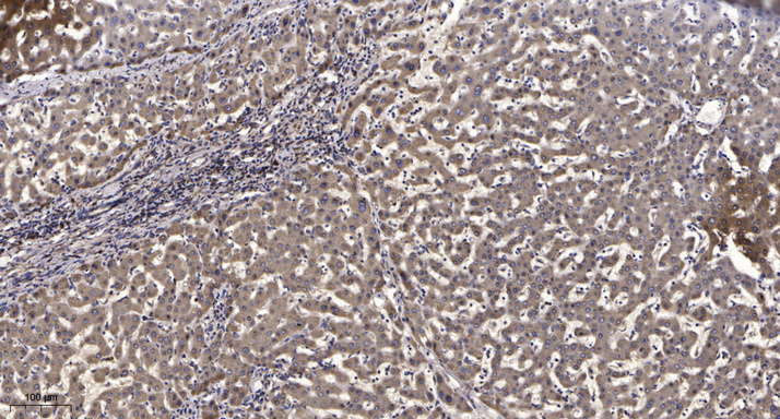
- Immunohistochemical analysis of paraffin-embedded human liver cancer. 1, Antibody was diluted at 1:200(4° overnight). 2, Tris-EDTA,pH9.0 was used for antigen retrieval. 3,Secondary antibody was diluted at 1:200(room temperature, 45min).


