PERK Polyclonal Antibody
- Catalog No.:YT3666
- Applications:IF;WB;IHC;ELISA
- Reactivity:Human;Mouse;Rat
- Target:
- PERK
- Fields:
- >>Mitophagy - animal;>>Autophagy - animal;>>Protein processing in endoplasmic reticulum;>>Apoptosis;>>Non-alcoholic fatty liver disease;>>Alzheimer disease;>>Parkinson disease;>>Amyotrophic lateral sclerosis;>>Prion disease;>>Pathways of neurodegeneration - multiple diseases;>>Hepatitis C;>>Measles;>>Herpes simplex virus 1 infection;>>Lipid and atherosclerosis
- Gene Name:
- EIF2AK3
- Protein Name:
- Eukaryotic translation initiation factor 2-alpha kinase 3
- Human Gene Id:
- 9451
- Human Swiss Prot No:
- Q9NZJ5
- Mouse Swiss Prot No:
- Q9Z2B5
- Rat Gene Id:
- 29702
- Rat Swiss Prot No:
- Q9Z1Z1
- Immunogen:
- The antiserum was produced against synthesized peptide derived from human EIF2AK3. AA range:947-996
- Specificity:
- PERK Polyclonal Antibody detects endogenous levels of PERK protein.
- Formulation:
- Liquid in PBS containing 50% glycerol, 0.5% BSA and 0.02% sodium azide.
- Source:
- Polyclonal, Rabbit,IgG
- Dilution:
- IF 1:50-200 WB 1:500 - 1:2000. IHC 1:100 - 1:300. ELISA: 1:40000. Not yet tested in other applications.
- Purification:
- The antibody was affinity-purified from rabbit antiserum by affinity-chromatography using epitope-specific immunogen.
- Concentration:
- 1 mg/ml
- Storage Stability:
- -15°C to -25°C/1 year(Do not lower than -25°C)
- Other Name:
- EIF2AK3;PEK;PERK;Eukaryotic translation initiation factor 2-alpha kinase 3;PRKR-like endoplasmic reticulum kinase;Pancreatic eIF2-alpha kinase;HsPEK
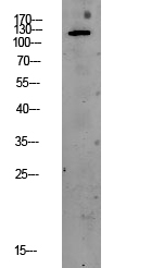
- Observed Band(KD):
- 125kD
- Background:
- The protein encoded by this gene phosphorylates the alpha subunit of eukaryotic translation-initiation factor 2, leading to its inactivation, and thus to a rapid reduction of translational initiation and repression of global protein synthesis. This protein is thought to modulate mitochondrial function. It is a type I membrane protein located in the endoplasmic reticulum (ER), where it is induced by ER stress caused by malfolded proteins. Mutations in this gene are associated with Wolcott-Rallison syndrome. [provided by RefSeq, Sep 2015],
- Function:
- catalytic activity:ATP + a protein = ADP + a phosphoprotein.,disease:Defects in EIF2AK3 are the cause of Wolcott-Rallison syndrome (WRS) [MIM:226980]; also known as multiple epiphyseal dysplasia with early-onset diabetes mellitus. WRS is a rare autosomal recessive disorder, characterized by permanent neonatal or early infancy insulin-dependent diabetes and, at a later age, epiphyseal dysplasia, osteoporosis, growth retardation and other multisystem manifestations, such as hepatic and renal dysfunctions, mental retardation and cardiovascular abnormalities.,domain:The lumenal domain senses perturbations in protein folding in the ER, probably through reversible interaction with HSPA5/BIP.,enzyme regulation:Perturbation in protein folding in the endoplasmic reticulum (ER) promotes reversible dissociation from HSPA5/BIP and oligomerization, resulting in transautophosphorylation and kinase act
- Subcellular Location:
- Endoplasmic reticulum membrane; Single-pass type I membrane protein.
- Expression:
- Ubiquitous. A high level expression is seen in secretory tissues.
NLRP3 inflammasome and endoplasmic reticulum stress in the epileptogenic zone in temporal lobe epilepsy: molecular insights into their interdependence. NEUROPATHOLOGY AND APPLIED NEUROBIOLOGY Neuropath Appl Neuro. 2020 Dec;46(7):770-785 WB,IF,IHC Mouse,Human 1:500,1:200,1:500 Temporal neocortices, bilateral hippocampi
Rce1 expression in renal cell carcinoma and its regulatory effect on 786-O cell apoptosis through endoplasmic reticulum stress. ACTA BIOCHIMICA ET BIOPHYSICA SINICA 2017 Mar 01 WB Human 1:2000 renal cancer tissues 786-O cell
Sodium fluoride induces apoptosis through reactive oxygen species-mediated endoplasmic reticulum stress pathway in Sertoli cells. Journal of Environmental Sciences 2015 Feb 24 WB Rat Sertoli cell
Activation of UPR Signaling Pathway is Associated With the Malignant Progression and Poor Prognosis in Prostate Cancer. PROSTATE 2016 Oct 08 IHC Human 1:100 Prostate cancer tissues
Cadmium induces liver dysfunction and ferroptosis through the endoplasmic stress-ferritinophagy axis ECOTOXICOLOGY AND ENVIRONMENTAL SAFETY Jiacheng Wu WB Mouse
α-Hederin induces human colorectal cancer cells apoptosis through disturbing protein homeostasis. CHEMICO-BIOLOGICAL INTERACTIONS Haibo Cheng WB Human HCT116 cell
- June 19-2018
- WESTERN IMMUNOBLOTTING PROTOCOL
- June 19-2018
- IMMUNOHISTOCHEMISTRY-PARAFFIN PROTOCOL
- June 19-2018
- IMMUNOFLUORESCENCE PROTOCOL
- September 08-2020
- FLOW-CYTOMEYRT-PROTOCOL
- May 20-2022
- Cell-Based ELISA│解您多样本WB检测之困扰
- July 13-2018
- CELL-BASED-ELISA-PROTOCOL-FOR-ACETYL-PROTEIN
- July 13-2018
- CELL-BASED-ELISA-PROTOCOL-FOR-PHOSPHO-PROTEIN
- July 13-2018
- Antibody-FAQs
- Products Images
- Immunofluorescence analysis of A549. 1,primary Antibody(red) was diluted at 1:200(4°C overnight). 2, Goat Anti Rabbit IgG (H&L) - Alexa Fluor 594 Secondary antibody was diluted at 1:1000(room temperature, 50min).3, Picture B: DAPI(blue) 10min.
- Immunofluorescence analysis of rat-lung tissue. 1,PERK Polyclonal Antibody(red) was diluted at 1:200(4°C,overnight). 2, Cy3 labled Secondary antibody was diluted at 1:300(room temperature, 50min).3, Picture B: DAPI(blue) 10min. Picture A:Target. Picture B: DAPI. Picture C: merge of A+B
- Immunofluorescence analysis of rat-spleen tissue. 1,PERK Polyclonal Antibody(red) was diluted at 1:200(4°C,overnight). 2, Cy3 labled Secondary antibody was diluted at 1:300(room temperature, 50min).3, Picture B: DAPI(blue) 10min. Picture A:Target. Picture B: DAPI. Picture C: merge of A+B
- Immunofluorescence analysis of mouse-lung tissue. 1,PERK Polyclonal Antibody(red) was diluted at 1:200(4°C,overnight). 2, Cy3 labled Secondary antibody was diluted at 1:300(room temperature, 50min).3, Picture B: DAPI(blue) 10min. Picture A:Target. Picture B: DAPI. Picture C: merge of A+B
- Immunohistochemical analysis of paraffin-embedded Human-Tonsil tissue. 1,PERK Polyclonal Antibody was diluted at 1:200(4°C,overnight). 2, Sodium citrate pH 6.0 was used for antibody retrieval(>98°C,20min). 3,Secondary antibody was diluted at 1:200(room tempeRature, 30min). Negative control was used by secondary antibody only.
- Immunohistochemical analysis of paraffin-embedded Human-stomach-cancer tissue. 1,PERK Polyclonal Antibody was diluted at 1:200(4°C,overnight). 2, Sodium citrate pH 6.0 was used for antibody retrieval(>98°C,20min). 3,Secondary antibody was diluted at 1:200(room tempeRature, 30min). Negative control was used by secondary antibody only.
- Immunohistochemical analysis of paraffin-embedded Human-Appendix tissue. 1,PERK Polyclonal Antibody was diluted at 1:200(4°C,overnight). 2, Sodium citrate pH 6.0 was used for antibody retrieval(>98°C,20min). 3,Secondary antibody was diluted at 1:200(room tempeRature, 30min). Negative control was used by secondary antibody only.
- Immunohistochemical analysis of paraffin-embedded Rat-heart tissue. 1,PERK Polyclonal Antibody was diluted at 1:200(4°C,overnight). 2, Sodium citrate pH 6.0 was used for antibody retrieval(>98°C,20min). 3,Secondary antibody was diluted at 1:200(room tempeRature, 30min). Negative control was used by secondary antibody only.
- Immunohistochemical analysis of paraffin-embedded Rat-liver tissue. 1,PERK Polyclonal Antibody was diluted at 1:200(4°C,overnight). 2, Sodium citrate pH 6.0 was used for antibody retrieval(>98°C,20min). 3,Secondary antibody was diluted at 1:200(room tempeRature, 30min). Negative control was used by secondary antibody only.
- Immunohistochemical analysis of paraffin-embedded Rat-kidney tissue. 1,PERK Polyclonal Antibody was diluted at 1:200(4°C,overnight). 2, Sodium citrate pH 6.0 was used for antibody retrieval(>98°C,20min). 3,Secondary antibody was diluted at 1:200(room tempeRature, 30min). Negative control was used by secondary antibody only.
- Immunohistochemical analysis of paraffin-embedded Mouse-liver tissue. 1,PERK Polyclonal Antibody was diluted at 1:200(4°C,overnight). 2, Sodium citrate pH 6.0 was used for antibody retrieval(>98°C,20min). 3,Secondary antibody was diluted at 1:200(room tempeRature, 30min). Negative control was used by secondary antibody only.
- Western blot analysis of lysates from MCF-7 cells, using PEK/PERK Antibody. The lane on the right is blocked with the synthesized peptide.