PI 3 kinase p85α/γ Polyclonal Antibody
- Catalog No.:YT3713
- Applications:WB;IHC;IF;ELISA
- Reactivity:Human;Mouse;Rat;Monkey;Chicken(testedbyourcustomer)
- Target:
- PI3 kinase p85α/γ
- Fields:
- >>EGFR tyrosine kinase inhibitor resistance;>>Endocrine resistance;>>Platinum drug resistance;>>ErbB signaling pathway;>>Ras signaling pathway;>>Rap1 signaling pathway;>>cAMP signaling pathway;>>Chemokine signaling pathway;>>HIF-1 signaling pathway;>>FoxO signaling pathway;>>Phosphatidylinositol signaling system;>>Sphingolipid signaling pathway;>>Phospholipase D signaling pathway;>>Autophagy - animal;>>mTOR signaling pathway;>>PI3K-Akt signaling pathway;>>AMPK signaling pathway;>>Apoptosis;>>Longevity regulating pathway;>>Longevity regulating pathway - multiple species;>>Cellular senescence;>>Axon guidance;>>VEGF signaling pathway;>>Osteoclast differentiation;>>Focal adhesion;>>Signaling pathways regulating pluripotency of stem cells;>>Platelet activation;>>Neutrophil extracellular trap formation;>>Toll-like receptor signaling pathway;>>C-type lectin receptor signaling pathway;>>JAK-STAT signaling pathway;>>Natural killer cell mediated cytotoxicity;>>T cell receptor signaling pathway;>
- Gene Name:
- PIK3R1/PIK3R3
- Protein Name:
- Phosphatidylinositol 3-kinase regulatory subunit alpha/gamma
- Human Gene Id:
- 5295/8503
- Human Swiss Prot No:
- P27986/Q92569
- Mouse Gene Id:
- 18708/18710
- Mouse Swiss Prot No:
- P26450
- Rat Gene Id:
- 25513/60664
- Rat Swiss Prot No:
- Q63787/Q63789
- Immunogen:
- The antiserum was produced against synthesized peptide derived from human PI3-kinase p85-alpha/gamma. AA range:436-485
- Specificity:
- PI 3-kinase p85α/γ Polyclonal Antibody detects endogenous levels of PI 3-kinase p85α/γ protein.
- Formulation:
- Liquid in PBS containing 50% glycerol, 0.5% BSA and 0.02% sodium azide.
- Source:
- Polyclonal, Rabbit,IgG
- Dilution:
- WB 1:500 - 1:2000. IHC 1:100 - 1:300. IF 1:200 - 1:1000. ELISA: 1:20000. Not yet tested in other applications.
- Purification:
- The antibody was affinity-purified from rabbit antiserum by affinity-chromatography using epitope-specific immunogen.
- Concentration:
- 1 mg/ml
- Storage Stability:
- -15°C to -25°C/1 year(Do not lower than -25°C)
- Other Name:
- PIK3R1;GRB1;Phosphatidylinositol 3-kinase regulatory subunit alpha;PI3-kinase regulatory subunit alpha;PI3K regulatory subunit alpha;PtdIns-3-kinase regulatory subunit alpha;Phosphatidylinositol 3-kinase 85 kDa regulatory subunit alpha;PI3-kinase subunit p85-alpha;PtdIns-3-kinase regulatory subunit p85-alpha
- Observed Band(KD):
- 54kD,83kD
- Background:
- Phosphatidylinositol 3-kinase phosphorylates the inositol ring of phosphatidylinositol at the 3-prime position. The enzyme comprises a 110 kD catalytic subunit and a regulatory subunit of either 85, 55, or 50 kD. This gene encodes the 85 kD regulatory subunit. Phosphatidylinositol 3-kinase plays an important role in the metabolic actions of insulin, and a mutation in this gene has been associated with insulin resistance. Alternative splicing of this gene results in four transcript variants encoding different isoforms. [provided by RefSeq, Jun 2011],
- Function:
- disease:Defects in PIK3R1 are a cause of severe insulin resistance.,domain:The SH3 domain mediates the binding to CBLB, and to HIV-1 Nef.,function:Binds to activated (phosphorylated) protein-Tyr kinases, through its SH2 domain, and acts as an adapter, mediating the association of the p110 catalytic unit to the plasma membrane. Necessary for the insulin-stimulated increase in glucose uptake and glycogen synthesis in insulin-sensitive tissues.,PTM:Polyubiquitinated in T-cells by CBLB; which does not promote proteasomal degradation but impairs association with CD28 and CD3Z upon T-cell activation.,similarity:Belongs to the PI3K p85 subunit family.,similarity:Contains 1 Rho-GAP domain.,similarity:Contains 1 SH3 domain.,similarity:Contains 2 SH2 domains.,subunit:Heterodimer of a p110 (catalytic) and a p85 (regulatory) subunits. Interacts with phosphorylated TOM1L1. Interacts with phosphorylat
- Subcellular Location:
- nucleus,cytoplasm,cis-Golgi network,cytosol,plasma membrane,cell-cell junction,phosphatidylinositol 3-kinase complex,phosphatidylinositol 3-kinase complex, class IA,membrane,perinuclear endoplasmic reticulum membrane,
- Expression:
- Isoform 2 is expressed in skeletal muscle and brain, and at lower levels in kidney and cardiac muscle. Isoform 2 and isoform 4 are present in skeletal muscle (at protein level).
Rapamycin Antagonizes BCRP-Mediated Drug Resistance Through the PI3K/Akt/mTOR Signaling Pathway in mPRα-Positive Breast Cancer. Frontiers in Oncology Front Oncol. 2021 Apr;0:352 WB Human 1:500 MDA-MB-231/BCRP cell,MDA-MB-453/BCRP cell
佟雪, et al. "基于 PI3K/AKT/mTOR 信号通路研究补肾法对体外培养小鼠卵母细胞成熟的促进作用." 北京中医药大学学报 44.6 (2021): 510-518.
恪雪, et al. "基于 PI3K/AKT/mTOR 信号通路研究补肾法对体外培养 小鼠卵母细胞成熟的促进作." Journal of Beijing University of Traditional Chinese Medicine 44.6 (2021).
- June 19-2018
- WESTERN IMMUNOBLOTTING PROTOCOL
- June 19-2018
- IMMUNOHISTOCHEMISTRY-PARAFFIN PROTOCOL
- June 19-2018
- IMMUNOFLUORESCENCE PROTOCOL
- September 08-2020
- FLOW-CYTOMEYRT-PROTOCOL
- May 20-2022
- Cell-Based ELISA│解您多样本WB检测之困扰
- July 13-2018
- CELL-BASED-ELISA-PROTOCOL-FOR-ACETYL-PROTEIN
- July 13-2018
- CELL-BASED-ELISA-PROTOCOL-FOR-PHOSPHO-PROTEIN
- July 13-2018
- Antibody-FAQs
- Products Images
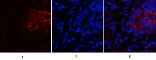
- Immunofluorescence analysis of human-lung tissue. 1,PI 3-kinase p85α/γ Polyclonal Antibody(red) was diluted at 1:200(4°C,overnight). 2, Cy3 labled Secondary antibody was diluted at 1:300(room temperature, 50min).3, Picture B: DAPI(blue) 10min. Picture A:Target. Picture B: DAPI. Picture C: merge of A+B
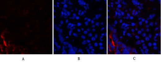
- Immunofluorescence analysis of rat-lung tissue. 1,PI 3-kinase p85α/γ Polyclonal Antibody(red) was diluted at 1:200(4°C,overnight). 2, Cy3 labled Secondary antibody was diluted at 1:300(room temperature, 50min).3, Picture B: DAPI(blue) 10min. Picture A:Target. Picture B: DAPI. Picture C: merge of A+B
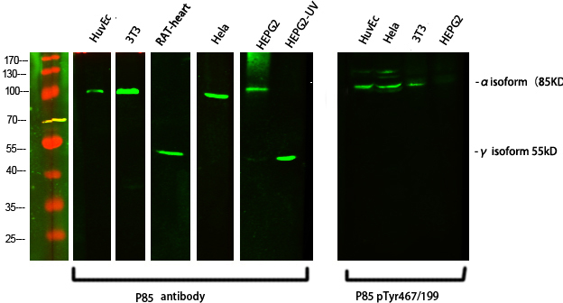
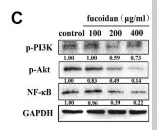
- Western Blot analysis of various cells using primary antibody diluted at 1:1000(4°C overnight). Secondary antibody:Goat Anti-rabbit IgG IRDye 800( diluted at 1:5000, 25°C, 1 hour)
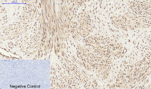
- Immunohistochemical analysis of paraffin-embedded Human-uterus tissue. 1,PI 3-kinase p85α/γ Polyclonal Antibody was diluted at 1:200(4°C,overnight). 2, Sodium citrate pH 6.0 was used for antibody retrieval(>98°C,20min). 3,Secondary antibody was diluted at 1:200(room tempeRature, 30min). Negative control was used by secondary antibody only.
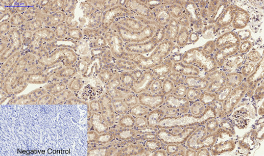
- Immunohistochemical analysis of paraffin-embedded Rat-kidney tissue. 1,PI 3-kinase p85α/γ Polyclonal Antibody was diluted at 1:200(4°C,overnight). 2, Sodium citrate pH 6.0 was used for antibody retrieval(>98°C,20min). 3,Secondary antibody was diluted at 1:200(room tempeRature, 30min). Negative control was used by secondary antibody only.
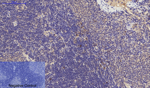
- Immunohistochemical analysis of paraffin-embedded Rat-spleen tissue. 1,PI 3-kinase p85α/γ Polyclonal Antibody was diluted at 1:200(4°C,overnight). 2, Sodium citrate pH 6.0 was used for antibody retrieval(>98°C,20min). 3,Secondary antibody was diluted at 1:200(room tempeRature, 30min). Negative control was used by secondary antibody only.
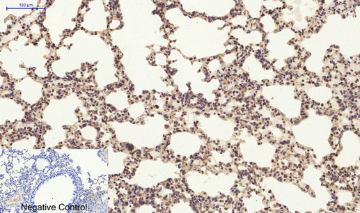
- Immunohistochemical analysis of paraffin-embedded Mouse-lung tissue. 1,PI 3-kinase p85α/γ Polyclonal Antibody was diluted at 1:200(4°C,overnight). 2, Sodium citrate pH 6.0 was used for antibody retrieval(>98°C,20min). 3,Secondary antibody was diluted at 1:200(room tempeRature, 30min). Negative control was used by secondary antibody only.

- Immunohistochemical analysis of paraffin-embedded Mouse-kidney tissue. 1,PI 3-kinase p85α/γ Polyclonal Antibody was diluted at 1:200(4°C,overnight). 2, Sodium citrate pH 6.0 was used for antibody retrieval(>98°C,20min). 3,Secondary antibody was diluted at 1:200(room tempeRature, 30min). Negative control was used by secondary antibody only.
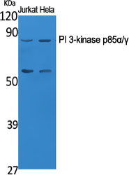
- Western Blot analysis of various cells using PI 3-kinase p85α/γ Polyclonal Antibody diluted at 1:1000
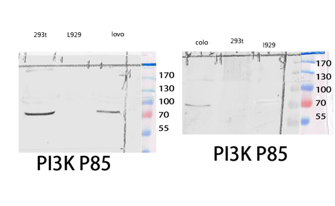
- Western blot analysis of 293T COLO lysis using PI 3-kinase p85α/γ antibody.
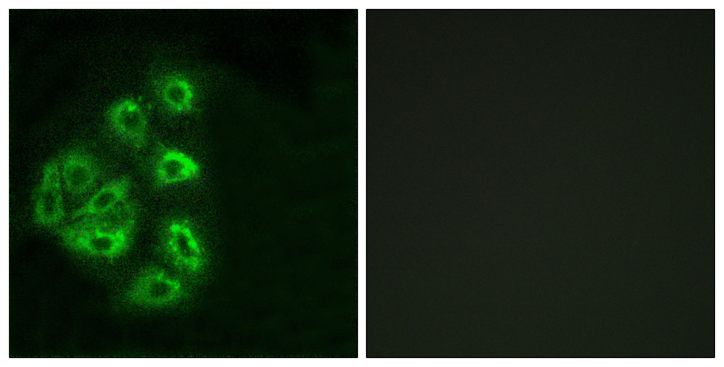
- Immunofluorescence analysis of HeLa cells, using PI3-kinase p85-alpha/gamma Antibody. The picture on the right is blocked with the synthesized peptide.
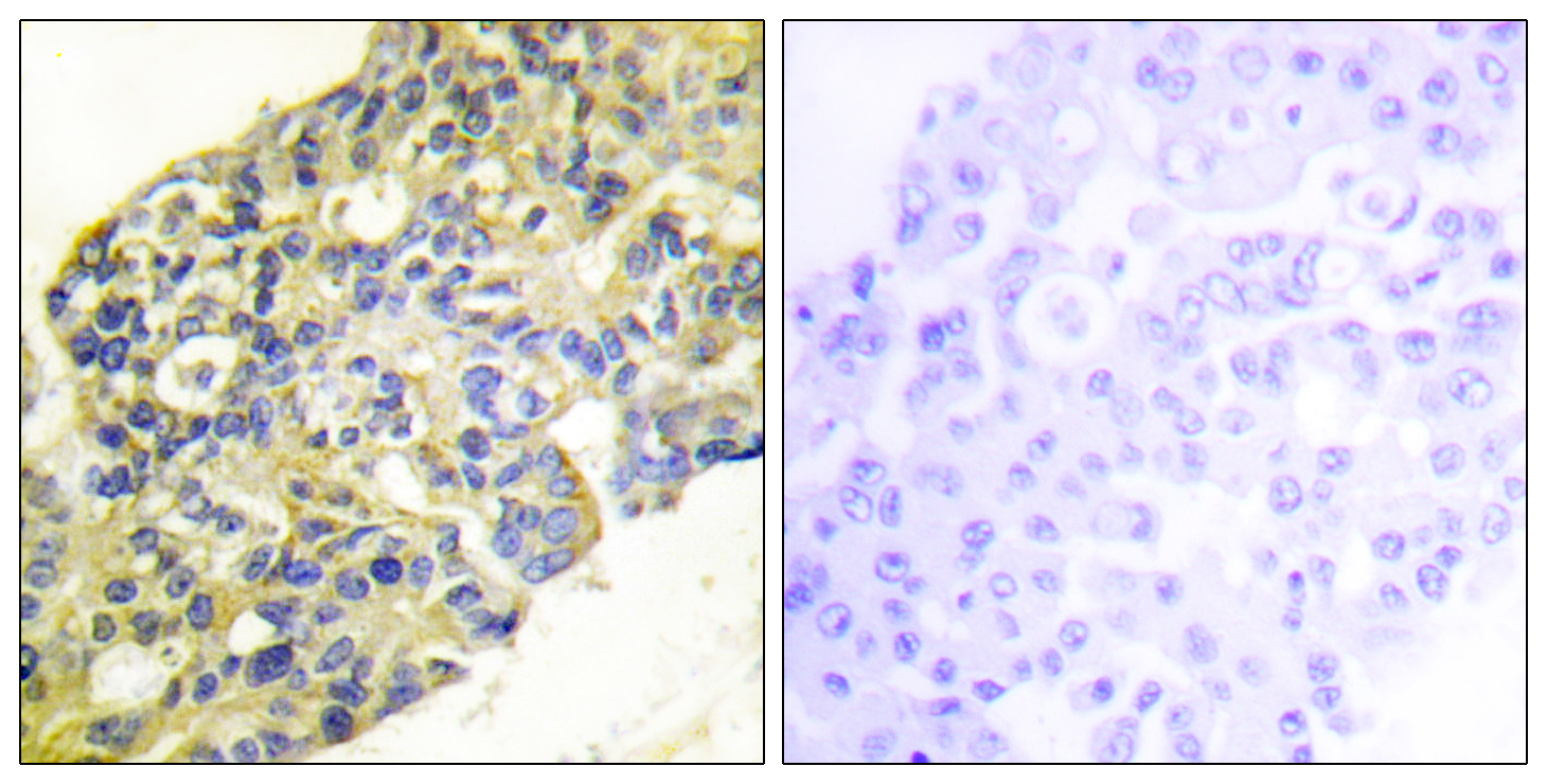
- Immunohistochemistry analysis of paraffin-embedded human breast carcinoma tissue, using PI3-kinase p85-alpha/gamma Antibody. The picture on the right is blocked with the synthesized peptide.
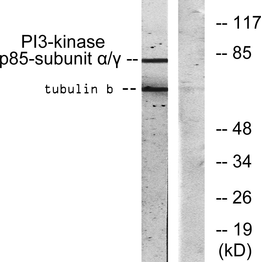
- Western blot analysis of lysates from COS7 cells, treated with H2O2 100uM 30', using PI3-kinase p85-alpha/gamma Antibody. The lane on the right is blocked with the synthesized peptide.