PIXβ Polyclonal Antibody
- Catalog No.:YT3741
- Applications:WB;IHC;IF;ELISA
- Reactivity:Human;Mouse;Rat
- Target:
- PIXβ
- Fields:
- >>Regulation of actin cytoskeleton;>>Yersinia infection
- Gene Name:
- ARHGEF7
- Protein Name:
- Rho guanine nucleotide exchange factor 7
- Human Gene Id:
- 8874
- Human Swiss Prot No:
- Q14155
- Mouse Gene Id:
- 54126
- Mouse Swiss Prot No:
- Q9ES28
- Rat Gene Id:
- 114559
- Rat Swiss Prot No:
- O55043
- Immunogen:
- The antiserum was produced against synthesized peptide derived from human ARHGEF7. AA range:311-360
- Specificity:
- PIXβ Polyclonal Antibody detects endogenous levels of PIXβ protein.
- Formulation:
- Liquid in PBS containing 50% glycerol, 0.5% BSA and 0.02% sodium azide.
- Source:
- Polyclonal, Rabbit,IgG
- Dilution:
- WB 1:500 - 1:2000. IHC 1:100 - 1:300. ELISA: 1:5000.. IF 1:50-200
- Purification:
- The antibody was affinity-purified from rabbit antiserum by affinity-chromatography using epitope-specific immunogen.
- Concentration:
- 1 mg/ml
- Storage Stability:
- -15°C to -25°C/1 year(Do not lower than -25°C)
- Other Name:
- ARHGEF7;COOL1;KIAA0142;P85SPR;PAK3BP;PIXB;Nbla10314;Rho guanine nucleotide exchange factor 7;Beta-Pix;COOL-1;PAK-interacting exchange factor beta;p85
- Observed Band(KD):
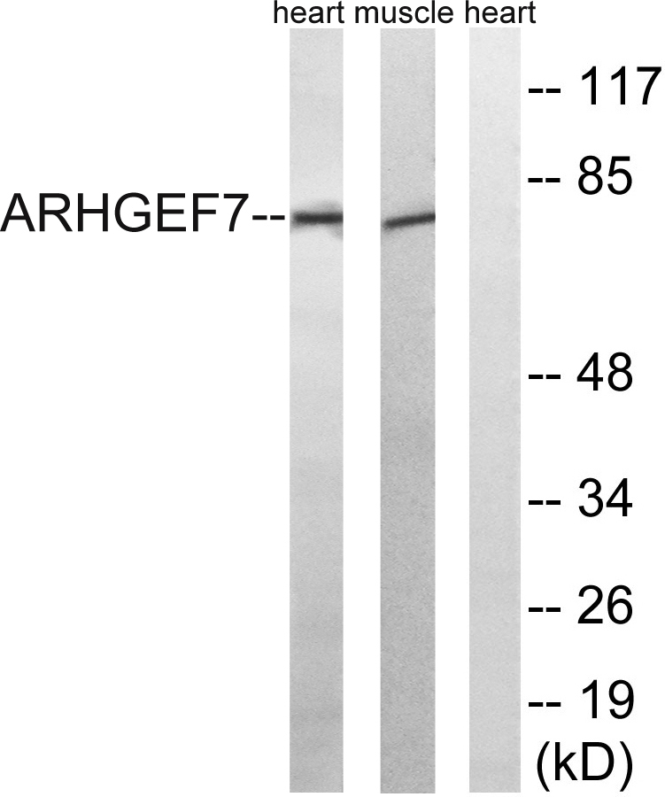
- 80kD
- Background:
- This gene encodes a protein that belongs to a family of cytoplasmic proteins that activate the Ras-like family of Rho proteins by exchanging bound GDP for GTP. It forms a complex with the small GTP binding protein Rac1 and recruits Rac1 to membrane ruffles and to focal adhesions. Multiple alternatively spliced transcript variants encoding different isoforms have been observed for this gene. [provided by RefSeq, Mar 2016],
- Function:
- function:Acts as a RAC1 guanine nucleotide exchange factor (GEF) and can induce membrane ruffling.,similarity:Contains 1 CH (calponin-homology) domain.,similarity:Contains 1 DH (DBL-homology) domain.,similarity:Contains 1 PH domain.,similarity:Contains 1 SH3 domain.,subunit:Interacts with PAK kinases through the SH3 domain. Interacts with GIT1 and TGFB1I1. Interacts with ITCH (By similarity). Interacts with unphosphorylated PAK1.,
- Subcellular Location:
- Cell junction, focal adhesion . Cell projection, ruffle . Cytoplasm, cell cortex . Cell projection, lamellipodium . Detected at cell adhesions. A small proportion is detected at focal adhesions.
- Expression:
- Amygdala,Bone marrow,Brain,Epithelium,Neuroblastoma,Testis,
- June 19-2018
- WESTERN IMMUNOBLOTTING PROTOCOL
- June 19-2018
- IMMUNOHISTOCHEMISTRY-PARAFFIN PROTOCOL
- June 19-2018
- IMMUNOFLUORESCENCE PROTOCOL
- September 08-2020
- FLOW-CYTOMEYRT-PROTOCOL
- May 20-2022
- Cell-Based ELISA│解您多样本WB检测之困扰
- July 13-2018
- CELL-BASED-ELISA-PROTOCOL-FOR-ACETYL-PROTEIN
- July 13-2018
- CELL-BASED-ELISA-PROTOCOL-FOR-PHOSPHO-PROTEIN
- July 13-2018
- Antibody-FAQs
- Products Images

- Western blot analysis of lysates from rat muscle and rat heart cells, using ARHGEF7 Antibody. The lane on the right is blocked with the synthesized peptide.

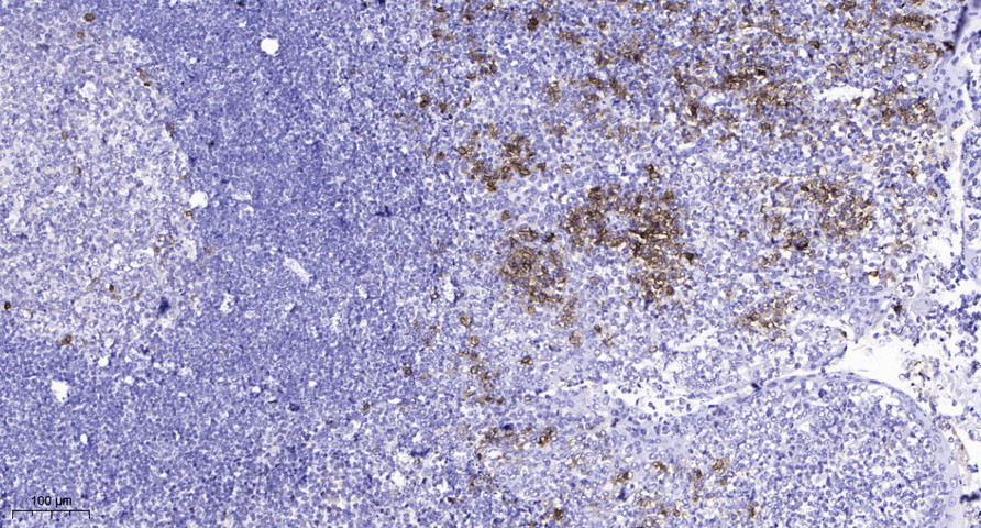
- Immunohistochemical analysis of paraffin-embedded human tonsil. 1, Antibody was diluted at 1:200(4° overnight). 2, Tris-EDTA,pH9.0 was used for antigen retrieval. 3,Secondary antibody was diluted at 1:200(room temperature, 45min).


