PKC δ Polyclonal Antibody
- Catalog No.:YT3761
- Applications:WB;IHC;IF;ELISA
- Reactivity:Human;Rat;Mouse;
- Target:
- PKC δ
- Fields:
- >>Chemokine signaling pathway;>>Autophagy - animal;>>Vascular smooth muscle contraction;>>NOD-like receptor signaling pathway;>>C-type lectin receptor signaling pathway;>>Fc gamma R-mediated phagocytosis;>>Neurotrophin signaling pathway;>>Inflammatory mediator regulation of TRP channels;>>GnRH signaling pathway;>>Estrogen signaling pathway;>>Type II diabetes mellitus;>>Insulin resistance;>>AGE-RAGE signaling pathway in diabetic complications;>>Prion disease;>>Shigellosis;>>Chemical carcinogenesis - reactive oxygen species;>>Diabetic cardiomyopathy
- Gene Name:
- PRKCD
- Protein Name:
- Protein kinase C delta type
- Human Gene Id:
- 5580
- Human Swiss Prot No:
- Q05655
- Mouse Swiss Prot No:
- P28867
- Immunogen:
- The antiserum was produced against synthesized peptide derived from human PKC delta. AA range:612-661
- Specificity:
- PKC δ Polyclonal Antibody detects endogenous levels of PKC δ protein.
- Formulation:
- Liquid in PBS containing 50% glycerol, 0.5% BSA and 0.02% sodium azide.
- Source:
- Polyclonal, Rabbit,IgG
- Dilution:
- WB 1:500 - 1:2000. IHC 1:100 - 1:300. ELISA: 1:10000.. IF 1:50-200
- Purification:
- The antibody was affinity-purified from rabbit antiserum by affinity-chromatography using epitope-specific immunogen.
- Concentration:
- 1 mg/ml
- Storage Stability:
- -15°C to -25°C/1 year(Do not lower than -25°C)
- Other Name:
- PRKCD;Protein kinase C delta type;Tyrosine-protein kinase PRKCD;nPKC-delta
- Observed Band(KD):
- 77kD
- Background:
- Protein kinase C (PKC) is a family of serine- and threonine-specific protein kinases that can be activated by calcium and the second messenger diacylglycerol. PKC family members phosphorylate a wide variety of protein targets and are known to be involved in diverse cellular signaling pathways. PKC family members also serve as major receptors for phorbol esters, a class of tumor promoters. Each member of the PKC family has a specific expression profile and is believed to play distinct roles in cells. The protein encoded by this gene is one of the PKC family members. Studies both in human and mice demonstrate that this kinase is involved in B cell signaling and in the regulation of growth, apoptosis, and differentiation of a variety of cell types. Alternatively spliced transcript variants encoding the same protein have been observed. [provided by RefSeq, Jul 2008],
- Function:
- catalytic activity:ATP + a protein = ADP + a phosphoprotein.,domain:The C1 domain, containing the phorbol ester/DAG-type region 1 (C1A) and 2 (C1B), is the diacylglycerol sensor.,domain:The C2 domain is a non-calcium binding domain. It binds proteins containing phosphotyrosine in a sequence-specific manner.,enzyme regulation:Three specific sites; Thr-507 (activation loop of the kinase domain), Ser-645 (turn motif) and Ser-664 (hydrophobic region), need to be phosphorylated for its full activation.,function:This is calcium-independent, phospholipid-dependent, serine- and threonine-specific enzyme. PKC is activated by diacylglycerol which in turn phosphorylates a range of cellular proteins. PKC also serves as the receptor for phorbol esters, a class of tumor promoters. May play a role in antigen-dependent control of B-cell function. Phosphorylates MUC1 in the C-terminal and regulates the i
- Subcellular Location:
- Cytoplasm . Cytoplasm, perinuclear region . Nucleus . Cell membrane ; Peripheral membrane protein . Mitochondrion . Endomembrane system . Translocates to the mitochondria upon apoptotic stimulation. Upon activation, translocates to the plasma membrane followed by partial location to the endolysosomes (PubMed:17303575). .
- Expression:
- Epithelium,Hippocampus,Liver,Platelet,Skin,
DBA-induced caspase-3-dependent apoptosis occurs through mitochondrial translocation of cyt-c in the rat hippocampus. Molecular BioSystems Mol Biosyst. 2017 Aug;13(9):1863-1873 WB Rat 1:1000 hippocampus
- June 19-2018
- WESTERN IMMUNOBLOTTING PROTOCOL
- June 19-2018
- IMMUNOHISTOCHEMISTRY-PARAFFIN PROTOCOL
- June 19-2018
- IMMUNOFLUORESCENCE PROTOCOL
- September 08-2020
- FLOW-CYTOMEYRT-PROTOCOL
- May 20-2022
- Cell-Based ELISA│解您多样本WB检测之困扰
- July 13-2018
- CELL-BASED-ELISA-PROTOCOL-FOR-ACETYL-PROTEIN
- July 13-2018
- CELL-BASED-ELISA-PROTOCOL-FOR-PHOSPHO-PROTEIN
- July 13-2018
- Antibody-FAQs
- Products Images

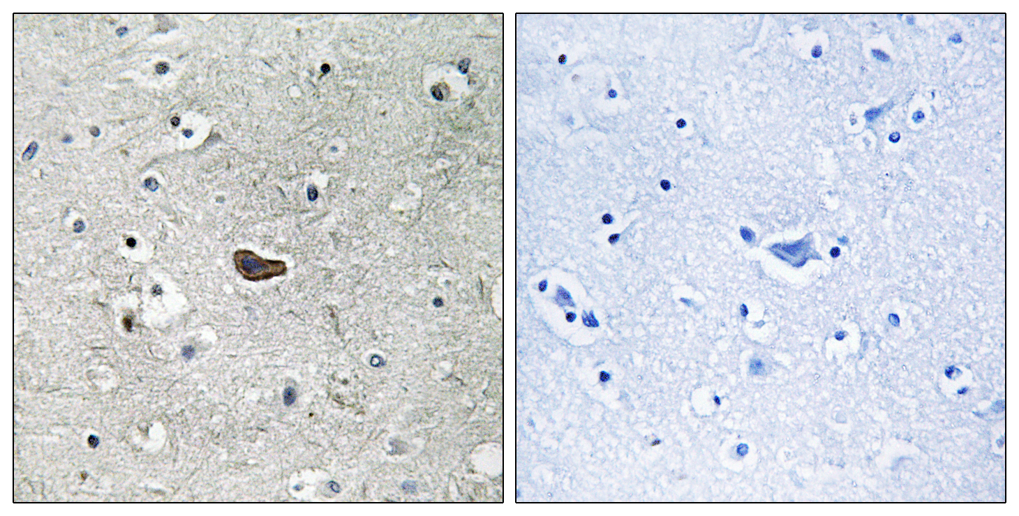
- Immunohistochemistry analysis of paraffin-embedded human brain tissue, using PKC delta Antibody. The picture on the right is blocked with the synthesized peptide.

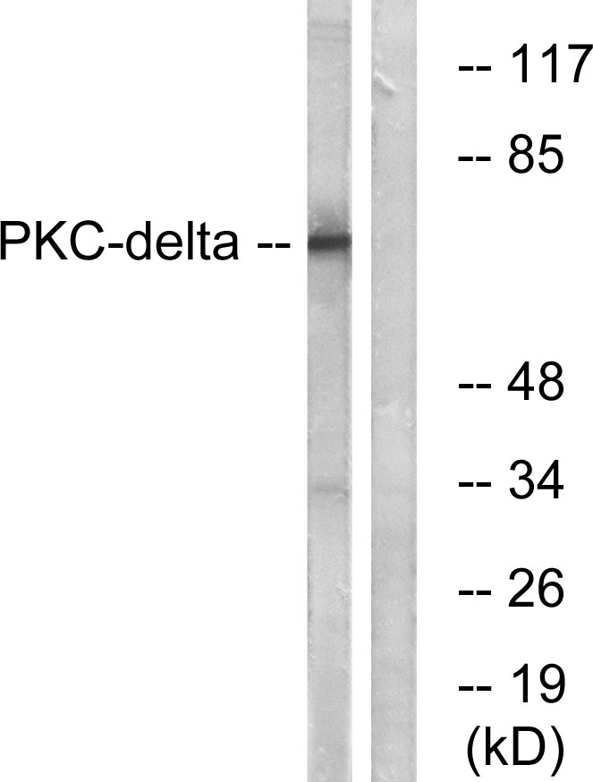
- Western blot analysis of lysates from MCF7 cells, using PKC delta Antibody. The lane on the right is blocked with the synthesized peptide.

