TACC1 Polyclonal Antibody
- Catalog No.:YT4521
- Applications:WB;IHC;IF;ELISA
- Reactivity:Human;Mouse
- Target:
- TACC1
- Gene Name:
- TACC1
- Protein Name:
- Transforming acidic coiled-coil-containing protein 1
- Human Gene Id:
- 6867
- Human Swiss Prot No:
- O75410
- Mouse Gene Id:
- 320165
- Mouse Swiss Prot No:
- Q6Y685
- Immunogen:
- The antiserum was produced against synthesized peptide derived from human TACC1. AA range:11-60
- Specificity:
- TACC1 Polyclonal Antibody detects endogenous levels of TACC1 protein.
- Formulation:
- Liquid in PBS containing 50% glycerol, 0.5% BSA and 0.02% sodium azide.
- Source:
- Polyclonal, Rabbit,IgG
- Dilution:
- WB 1:500 - 1:2000. IHC 1:100 - 1:300. IF 1:200 - 1:1000. ELISA: 1:10000. Not yet tested in other applications.
- Purification:
- The antibody was affinity-purified from rabbit antiserum by affinity-chromatography using epitope-specific immunogen.
- Concentration:
- 1 mg/ml
- Storage Stability:
- -15°C to -25°C/1 year(Do not lower than -25°C)
- Other Name:
- TACC1;KIAA1103;Transforming acidic coiled-coil-containing protein 1;Gastric cancer antigen Ga55;Taxin-1
- Observed Band(KD):
- 87kD
- Background:
- This locus may represent a breast cancer candidate gene. It is located close to FGFR1 on a region of chromosome 8 that is amplified in some breast cancers. Three transcript variants encoding different isoforms have been found for this gene. [provided by RefSeq, Apr 2009],
- Function:
- alternative products:Additional isoforms seem to exist,developmental stage:Expressed at high level during early embryogenesis.,function:Likely involved in the processes that promote cell division prior to the formation of differentiated tissues.,miscellaneous:Down-regulated in a subset of cases of breast cancer.,PTM:Isoform 1 is heavily phosphorylated; isoform 6 is not. Phosphorylated upon DNA damage, probably by ATM or ATR.,similarity:Belongs to the TACC family.,similarity:Contains 2 SPAZ (Ser/Pro-rich AZU-1) domains.,subcellular location:Nucleus during interphase. Weakly concentrated at centrosomes during mitosis.,subunit:Interacts with KIAA0097/CH-TOG and with the oncogenic transcription factor YEATS4. Interacts with the Aurora kinases A and B (STK6 and AURKB). Interacts with LSM7, TDRD7 and SNRPG. Interacts with GCN5L2 and PCAF.,tissue specificity:Isoform 1, isoform 3 and isoform 5 a
- Subcellular Location:
- Cytoplasm . Nucleus . Cytoplasm, cytoskeleton, microtubule organizing center, centrosome . Midbody . Nucleus during interphase. Weakly concentrated at centrosomes during mitosis and colocalizes with AURKC at the midbody during cytokinesis. .; [Isoform 5]: Membrane ; Lipid-anchor .; [Isoform 10]: Cytoplasm .
- Expression:
- Isoform 1, isoform 3 and isoform 5 are ubiquitous. Isoform 2 is strongly expressed in the brain, weakly detectable in lung and colon, and overexpressed in gastric cancer. Isoform 4 is not detected in normal tissues, but strong expression was found in gastric cancer tissues. Down-regulated in a subset of cases of breast cancer.
- June 19-2018
- WESTERN IMMUNOBLOTTING PROTOCOL
- June 19-2018
- IMMUNOHISTOCHEMISTRY-PARAFFIN PROTOCOL
- June 19-2018
- IMMUNOFLUORESCENCE PROTOCOL
- September 08-2020
- FLOW-CYTOMEYRT-PROTOCOL
- May 20-2022
- Cell-Based ELISA│解您多样本WB检测之困扰
- July 13-2018
- CELL-BASED-ELISA-PROTOCOL-FOR-ACETYL-PROTEIN
- July 13-2018
- CELL-BASED-ELISA-PROTOCOL-FOR-PHOSPHO-PROTEIN
- July 13-2018
- Antibody-FAQs
- Products Images
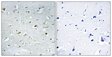
- Immunohistochemical analysis of paraffin-embedded Human brain. Antibody was diluted at 1:100(4° overnight). High-pressure and temperature Tris-EDTA,pH8.0 was used for antigen retrieval. Negetive contrl (right) obtaned from antibody was pre-absorbed by immunogen peptide.
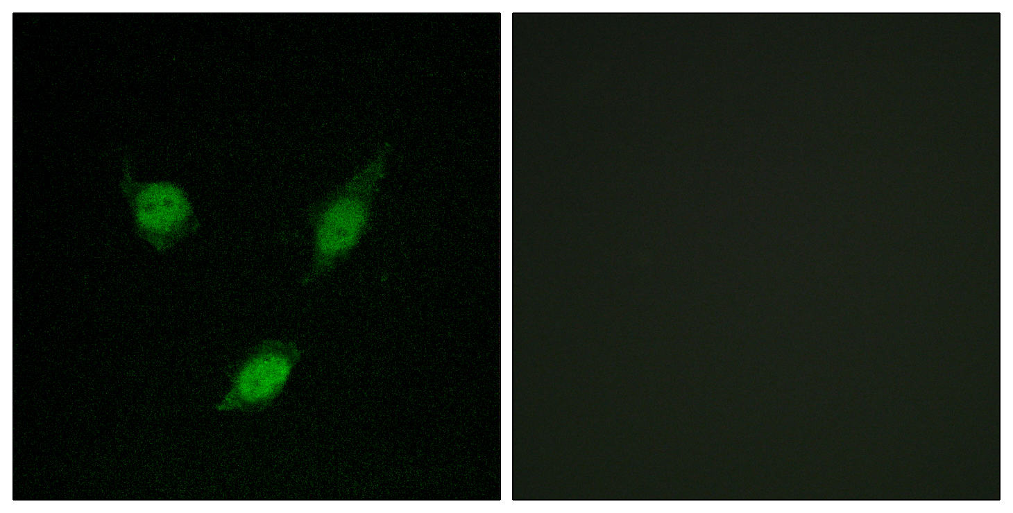
- Immunofluorescence analysis of MCF7 cells, using TACC1 Antibody. The picture on the right is blocked with the synthesized peptide.
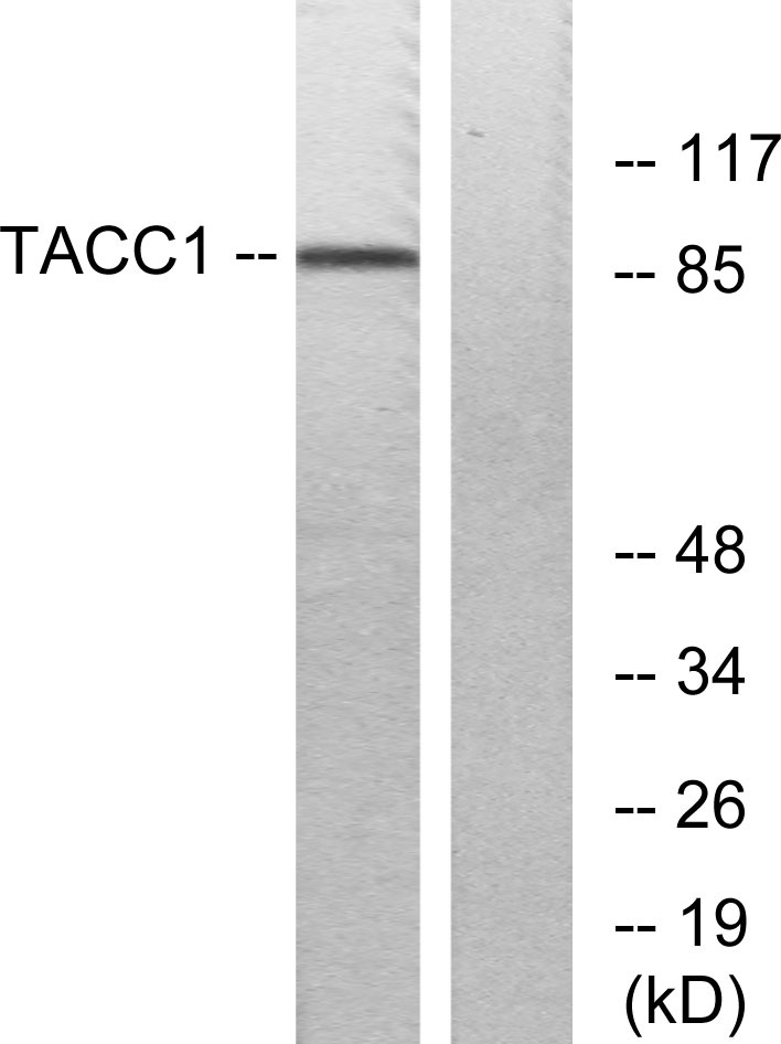
- Western blot analysis of lysates from K562 cells, using TACC1 Antibody. The lane on the right is blocked with the synthesized peptide.
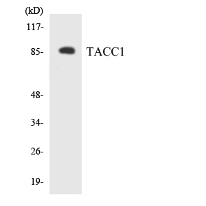
- Western blot analysis of the lysates from HeLa cells using TACC1 antibody.


